Every implant-based reconstruction is a bet on the mastectomy flaps. Mastectomy skin flap necrosis is the complication that turns good operations bad: it exposes devices, delays adjuvant therapy and undoes envelopes that can never be fully rebuilt. This professional overview covers the risk factors, the assessment tools including indocyanine green angiography, and the intraoperative decisions that flap quality should drive.

Why flaps fail
The mastectomy flap survives on the subdermal plexus. Necrosis follows when that plexus is too thin, too damaged or too stretched: overly aggressive dissection, flaps thinned past the oncologically necessary plane, excessive tension from an over-ambitious implant, and systemic factors that compromise microcirculation. The recurring patient-level risk factors across published series are smoking, high BMI, diabetes, prior irradiation and large breast size; the recurring surgical factors are flap thickness, incision choice and device volume.
Clinical assessment and its limits
Experienced surgeons read flaps continuously: colour, capillary refill, dermal bleeding from the cut edge, warmth. Clinical judgement, however, is documented to overestimate perfusion in a meaningful fraction of cases, particularly in the pale, adrenaline-infiltrated field of a fresh mastectomy. That gap between how a flap looks and how it is perfused is exactly where objective tools earn their place.
Indocyanine green angiography
Intraoperative ICG angiography injects a fluorescent dye and images its arrival in the flap in real time, mapping perfusion while decisions can still be changed. Studies and systematic reviews report that ICG-guided assessment identifies poorly perfused zones more reliably than clinical examination and is associated with reduced necrosis rates when its findings actually change management: trimming compromised edges, reducing implant volume, or converting a planned direct-to-implant into a staged expander reconstruction. The tool has costs and false positives, and protocols vary; its value is greatest exactly where the stakes are highest, in nipple-sparing mastectomy and single-stage reconstruction.
Decisions flap quality should drive
- Reconstruction timing. Robust flaps permit immediate reconstruction; marginal flaps argue for an expander at low fill, or in extremes, delayed reconstruction. This is the consensus framework: keep every option open until the flaps declare themselves.
- Device volume. The implant serves the envelope, never the reverse; tension is the enemy of the plexus.
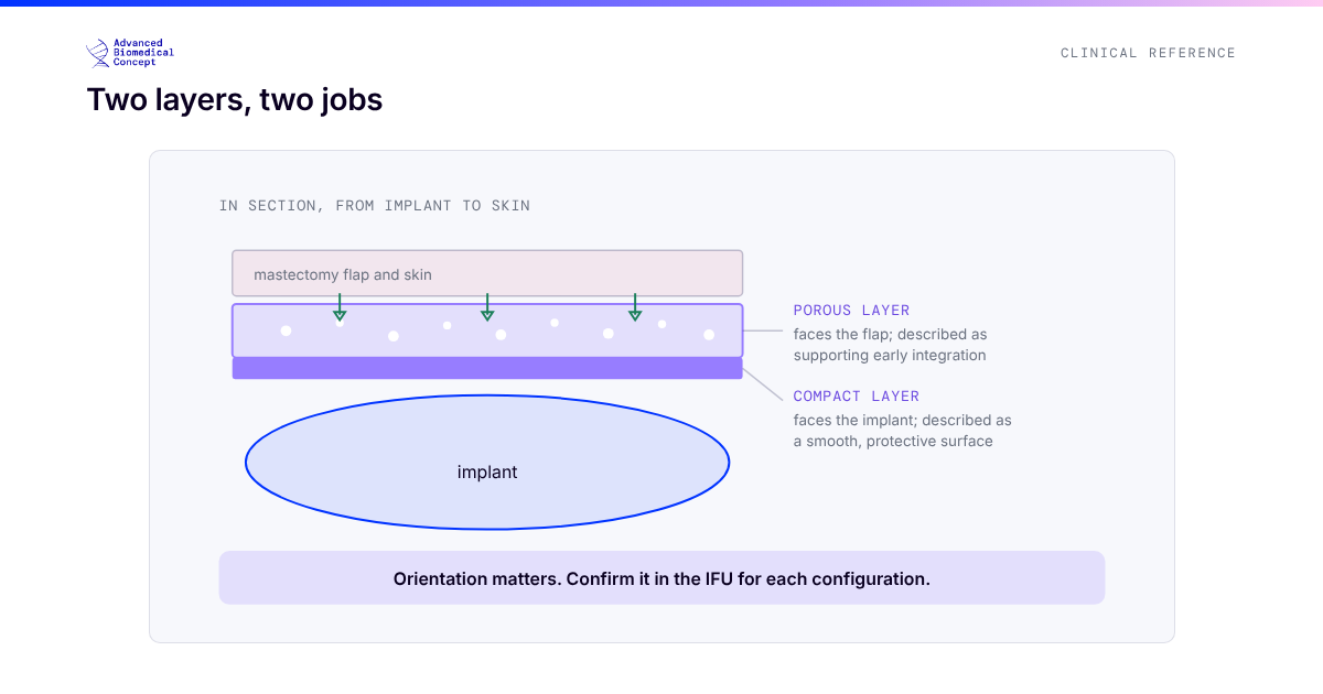
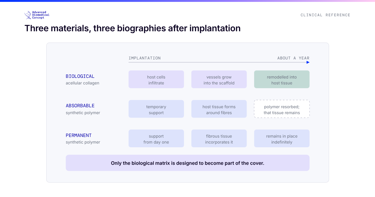
- Plane and support. Where flaps are adequate but the patient is slim, prepectoral placement with a biological matrix and planned fat grafting manages the thin-cover problem without recruiting muscle.
When necrosis happens anyway
Management is graded to depth and device exposure: superficial epidermolysis is dressed and watched; full-thickness necrosis over an implant is debrided early, because the race between granulation and device exposure is rarely won by waiting. Salvage rates are meaningfully better when intervention is early and the pocket remains uncontaminated. Every unit’s protocol differs in detail; the shared principle is that necrosis declared early and managed actively costs an edge, while necrosis denied costs the reconstruction.
The bottom line
Flap perfusion is the single variable that should shape every intraoperative reconstruction decision. Assess it honestly, augment judgement with objective imaging where available, and let the flaps choose the operation. The reconstructions that fail are rarely the ones where the team was too cautious.
Key published sources
- Considerations for patient selection: prepectoral versus subpectoral implant-based reconstruction. Gland Surgery. 2019.
- Highton L, et al. Prepectoral implant-based breast reconstruction: a joint consensus guide. ecancermedicalscience. 2019.
- Systematic reviews of indocyanine green angiography in mastectomy flap assessment, plastic and reconstructive surgery literature, 2018 onward.
Important information This article is general education, not medical advice. Tap to read the full medical, regulatory & legal notice.
This article is provided by Advanced Biomedical Concept for general informational and educational purposes only. It does not constitute medical, surgical, diagnostic, or other professional healthcare advice, and it is not a substitute for consultation with a suitably qualified healthcare professional. Reading it creates no doctor–patient or other professional relationship. Nothing here should be used to diagnose, treat, cure, or prevent any disease or condition, or to make decisions about medical care. Always seek the advice of a physician or other qualified provider with any questions about a medical condition or treatment, and never disregard or delay professional advice because of something you have read here.
Any reference to Advanced Biomedical Concept products (including ExaShape and ExaFat) is provided for general information only. These are medical devices intended to be used by appropriately trained healthcare professionals strictly in accordance with their applicable, current Instructions for Use (IFU). Regulatory status (including CE marking and classification under the EU Medical Device Regulation), market availability, and approved indications differ between countries and may change over time; a device referred to here may not be available or approved in your country. This content is not intended as, and must not be relied upon as, a statement or claim of safety, clinical performance, or efficacy beyond what is set out in the applicable IFU and current regulatory approvals. Product-specific information is directed at healthcare professionals and is not intended to promote, advertise, or offer any medical device to the general public where such promotion is restricted or prohibited by law.
Statements reflect information believed to be accurate at the date of publication and may be updated or corrected without notice. Any statements regarding the future are expectations or general views, not guarantees. Clinical outcomes depend on many individual factors, results vary from person to person, and no particular result is promised or implied. Where third-party studies, sources, or external links are referenced, they are provided for convenience and information only and do not imply endorsement, and Advanced Biomedical Concept is not responsible for their content.
To the fullest extent permitted by applicable law, Advanced Biomedical Concept and its officers, employees, contributors, and representatives (each acting in that capacity and on behalf of Advanced Biomedical Concept) accept no liability for any loss, injury, or damage of any kind arising directly or indirectly from use of, or reliance on, this content. Nothing in this notice excludes or limits any liability that cannot lawfully be excluded or limited.
Questions about this material can be directed to us via our contact page.
Last reviewed: August 24, 2026.