The evidence articles on this site argue about planes, stages and materials. This one is practical. It is written for the surgeon who has decided that a biological support material belongs in the reconstruction and wants to know four things: why ExaShape®, in which cases, which configuration, and how it is used in theatre. Where this article describes handling, it describes principles; the Instructions for Use are the authority on product-specific steps, and nothing here replaces them.
Why use a support material at all
In the prepectoral plane, the implant has nothing to hold it except the mastectomy flap and whatever is placed around it. A support material defines the pocket, controls implant position and the lower pole, and stands between the device and a flap that may be only a few millimetres thick. That is why almost every published prepectoral series uses one, and why the question in that plane is which material rather than whether. The evidence behind that is set out in meshes in breast reconstruction and prepectoral versus subpectoral: the evidence.
In the subpectoral and dual planes, a support material extends the released muscle over the lower pole. In two-stage reconstruction it supports the expander pocket during expansion. Each of those jobs has an ExaShape configuration.
What ExaShape is
ExaShape® is an acellular bovine pericardium membrane, CE marked under EU MDR 2017/745. Pericardium is a naturally layered collagen tissue, thinner and lower in biological mass than dermis, and ExaShape keeps that structure as a bilayer: a compact layer and a porous layer with different jobs.
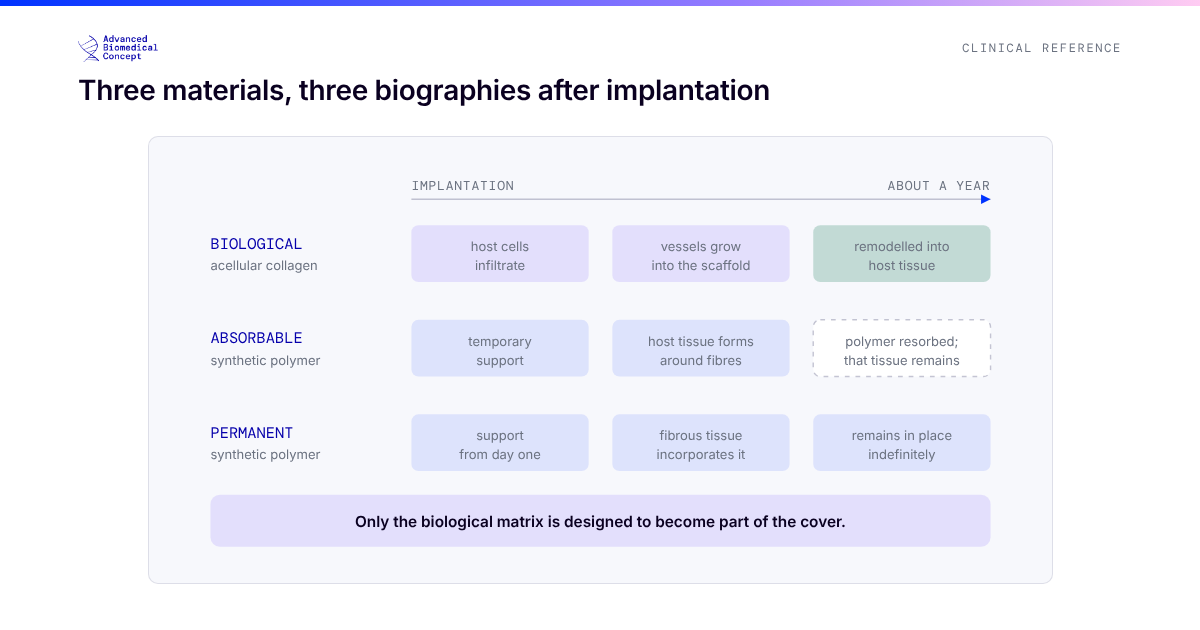
Why does a thinner, lower-mass biological material matter? Because the two recognised weaknesses of biological matrices in the evidence are seroma and the time it takes for a thick sheet of collagen to revascularise under a thin flap. A material with less mass and more porosity is designed to address both, and preclinical work with acellular pericardial meshes around breast implants in rats showed reduced capsular myofibroblast markers and inflammation and increased capillary density, with remodelling influenced by the product’s structure.1 That is mechanism, not clinical proof, and it is the rationale for the design.
Which configuration, for which plan
The ExaShape line is a set of configurations rather than a single sheet, because the job a support material does depends on the plane and the stage. The choice follows from the reconstructive plan.
In which cases surgeons use it
The configurations map onto recognisable clinical situations. These are the ones most often discussed by surgeons using bovine pericardium; the indication for any individual patient remains the surgeon’s judgement within the IFU.
Nipple-sparing or skin-sparing mastectomy with a good flap, planned direct-to-implant. The core use. The pocket is wrapped around the implant and the wrapped implant is placed prepectorally in a single stage. This is the setting of the largest published ExaShape-type series: 65 DTI prepectoral reconstructions with a bovine pericardium pocket across several centres, mean follow-up 21.3 months, with minor complications in four breasts, major complications in two, and no significant capsular contracture.2
Risk-reducing bilateral mastectomy. Often the ideal patient: young, healthy tissue, no radiotherapy planned, and a strong preference for one operation. The case for prepectoral DTI with a pocket is at its strongest here.
Larger implants or a preference for minimal handling. The pre-shaped 3D pocket covers larger implants and is designed for a full no-touch wrap, reducing handling of the device.
Equivocal flap on the day. If the flap will not carry a full-volume implant, the plan converts to two stages. The Expander configuration supports the expander pocket during expansion, keeping the same material through to the exchange.
Subpectoral or dual-plane reconstruction. Where the surgeon prefers or the patient needs muscle cover, Grid extends the released pectoralis over the lower pole.
Thin flaps. Here a biological material has its clearest rationale, because it is designed to become part of the cover. In a series selected for prepectoral patients with flaps thinner than 1 cm, surgeons wrapped the implant in bovine pericardium specifically for this reason, reporting no major complications and rippling in a quarter of cases, corrected by secondary fat grafting.3 Thin flaps also call for the most careful selection: if perfusion is in doubt, two stages are safer than one.
How it is used: the theatre sequence
Decide on the flap, not the plan. The mastectomy flap is assessed after the mastectomy is complete and with the patient warm and normotensive. If it will not carry a full-volume implant, convert to two stages. That decision is covered in detail in reading the mastectomy flap.
Wrap on the back table. The pocket design allows the implant to be wrapped away from the patient. In the multicentre pocket series, wrapping was done on a back table at the same time as haemostasis, drain positioning and preparation of the field, and mean total operating time was 1.42 hours.2
Handle the device as little as possible. Change gloves before handling the implant, keep exposure short, irrigate the pocket, and insert the wrapped implant in a single movement. The 3D pocket is designed for a no-touch technique for exactly this reason.
Respect orientation. Place the porous side towards the tissue that will integrate it and the compact side towards the device, as the IFU specifies for each configuration.
Plan for fluid. Seroma is the complication biological matrices are most often criticised for. Drains, a drain-removal protocol, and attention to dead space matter as much as the material; in one dermal matrix series, a change in fixation technique alone was associated with a fall in seroma from 13.9% to 2.3%.4
Plan for refinement. Prepectoral reconstruction in thinner patients often benefits from a later round of fat grafting to soften the upper pole and correct rippling. In the pocket series, 7 patients had fat grafting at six months for that purpose.2 A consistent, well-processed graft makes that step more predictable; the EXAFAT® closed-loop system, from the same manufacturer, was designed for breast fat grafting and is the natural partner for the refinement stage. Graft planning is covered in planning fat grafting across sessions.
Ideas worth discussing with your team
Surgeons who use ExaShape regularly tend to raise the same practical questions. A few worth considering when setting up a pathway:
Make selection explicit. Agree in advance which preoperative factors make a patient a prepectoral DTI candidate and which mean two stages from the start, and record the intraoperative flap decision. Units that do this find it easier to audit their outcomes and to explain their conversion rate.
Audit the first cases. Every published technique series shows a learning curve, and the MGH DTI experience saw complications fall by half after the first year.5 Prospective audit of the first cases, including seroma and drain duration, is the fastest way through it.
Build the refinement stage into the consent. A short fat grafting procedure is much easier to discuss at the outset, as an expected step, than to introduce later as a surprise.
Use the training. ABC’s e-learning platform and hands-on training in Rome cover configuration choice, wrapping and insertion in more detail than a web page can.
The short version
Use ExaShape where a biological material has a job to do: forming the prepectoral pocket, extending the muscle, or supporting an expander. Choose the configuration by that job. Decide on the flap, wrap on the back table, handle the device as little as possible, plan for fluid, and plan for refinement. The evidence for the material class is consistent and growing; the evidence for any individual patient is what the flap looks like on the day.
References
- Bernardini R, Varvaras D, D’Amico F, et al. Biological acellular pericardial mesh regulated tissue integration and remodeling in a rat model of breast prosthetic implantation. J Biomed Mater Res B Appl Biomater. 2020;108(2):577-590. doi:10.1002/jbm.b.34413
- De Vita R, Mangialardi ML, Pozzi M, et al. A pericardium bovine matrix pocket in DTI prepectoral breast reconstruction. Clin Breast Cancer. 2024;24(7):e613-e621. doi:10.1016/j.clbc.2024.06.004. Disclosure: the first author is a consultant for ExaShape.
- Casella D, Rocco N, Luridiana G, et al. Enhancing breast reconstruction with bovine pericardium: a preliminary STEP (Surgical Techniques and Efficacy in Pericardium use) towards improved outcomes. J Clin Med. 2025;14(17):6296. doi:10.3390/jcm14176296. This series used a different bovine pericardium product.
- Tettamanzi M, Arrica G, Ziani F, et al. Prepectoral breast reconstruction with prosthesis and acellular dermal matrix: a new technique of ADM implantation and fixation. Plast Reconstr Surg Glob Open. 2024;12(6):e5895. doi:10.1097/GOX.0000000000005895
- Colwell AS, Damjanovic B, Zahedi B, Medford-Davis L, Hertl C, Austen WG. Retrospective review of 331 consecutive immediate single-stage implant reconstructions with acellular dermal matrix: indications, complications, trends, and costs. Plast Reconstr Surg. 2011;128(6):1170-1178. doi:10.1097/PRS.0b013e318230c2f6
This article is educational material for healthcare professionals and does not replace the Instructions for Use. Product configurations, sizes, indications, preparation and handling must be confirmed in the current ExaShape® IFU. No clinical superiority over other materials is claimed. ExaShape® and EXAFAT® are CE marked per EU MDR 2017/745 and are for professional use only.
Important information This article is general education, not medical advice. Tap to read the full medical, regulatory & legal notice.
This article is provided by Advanced Biomedical Concept for general informational and educational purposes only. It does not constitute medical, surgical, diagnostic, or other professional healthcare advice, and it is not a substitute for consultation with a suitably qualified healthcare professional. Reading it creates no doctor–patient or other professional relationship. Nothing here should be used to diagnose, treat, cure, or prevent any disease or condition, or to make decisions about medical care. Always seek the advice of a physician or other qualified provider with any questions about a medical condition or treatment, and never disregard or delay professional advice because of something you have read here.
Any reference to Advanced Biomedical Concept products (including ExaShape and ExaFat) is provided for general information only. These are medical devices intended to be used by appropriately trained healthcare professionals strictly in accordance with their applicable, current Instructions for Use (IFU). Regulatory status (including CE marking and classification under the EU Medical Device Regulation), market availability, and approved indications differ between countries and may change over time; a device referred to here may not be available or approved in your country. This content is not intended as, and must not be relied upon as, a statement or claim of safety, clinical performance, or efficacy beyond what is set out in the applicable IFU and current regulatory approvals. Product-specific information is directed at healthcare professionals and is not intended to promote, advertise, or offer any medical device to the general public where such promotion is restricted or prohibited by law.
Statements reflect information believed to be accurate at the date of publication and may be updated or corrected without notice. Any statements regarding the future are expectations or general views, not guarantees. Clinical outcomes depend on many individual factors, results vary from person to person, and no particular result is promised or implied. Where third-party studies, sources, or external links are referenced, they are provided for convenience and information only and do not imply endorsement, and Advanced Biomedical Concept is not responsible for their content.
To the fullest extent permitted by applicable law, Advanced Biomedical Concept and its officers, employees, contributors, and representatives (each acting in that capacity and on behalf of Advanced Biomedical Concept) accept no liability for any loss, injury, or damage of any kind arising directly or indirectly from use of, or reliance on, this content. Nothing in this notice excludes or limits any liability that cannot lawfully be excluded or limited.
Questions about this material can be directed to us via our contact page.
Last reviewed: October 15, 2026.