Prepectoral reconstruction went from a niche technique to a mainstream one in under a decade. In the UK iBRA cohort, recruited between 2014 and 2016, just 42 of 2,108 patients (2%) had prepectoral implants; most had subpectoral reconstruction supported by biological or synthetic mesh.1 A few years later, prepectoral placement was routine in many units. Practice moved faster than evidence usually does, which makes it worth asking precisely what the evidence now says, how strong it is, and what it still cannot answer.
The mechanics of the two planes, what changes when the implant moves in front of the muscle, are covered in plane selection in direct-to-implant reconstruction. This article is about the data.
The early signal
One of the first comparative series came from UCSF. Sbitany and colleagues compared 84 breasts reconstructed prepectorally with complete matrix coverage against 186 reconstructed in a partial submuscular, dual-plane position after nipple-sparing mastectomy. Overall complication rates were essentially identical, 17.9% against 18.8%, and the authors reported significantly less acute and chronic pain and animation deformity in the prepectoral group. On that basis, prepectoral reconstruction became their technique for all oncologically suitable nipple-sparing patients.2
The finding that made the technique credible was the first one: equivalent safety. The benefits on pain and animation were expected from the anatomy. What surgeons needed to know was that removing the muscle from the construct did not make it more dangerous.
The meta-analysis
By 2023 enough comparative studies existed to pool. Ostapenko and colleagues included 15 studies and 3,101 patients published between 2011 and 2021. Compared with subpectoral reconstruction, prepectoral reconstruction was associated with less capsular contracture, much less animation deformity, and less prosthesis failure. It was not associated with any significant difference in overall complications, seroma, haematoma, infection, skin flap necrosis, local recurrence or BREAST-Q scores.3
Three things in that result deserve emphasis. The animation finding is not a statistical nuance; an odds ratio of 0.02 means animation deformity is, for practical purposes, a feature of the subpectoral plane. The capsular contracture finding is clinically important because contracture is a late complication that drives revision years after the index operation. And the prosthesis failure finding answers the early fear about the technique directly: removing the muscle did not make implant loss more likely; the pooled data suggest the opposite.
How strong is this evidence?
Honestly: moderate. The meta-analysis pooled observational studies, not randomised trials, and that limits what it can establish.
The central problem is selection. Surgeons choose prepectoral placement for patients with good flaps, and good flaps do well in any plane. Some of the advantage seen in observational data may therefore reflect who was selected rather than where the implant was placed. The UK Best-BRA trial was designed to address exactly this: a pilot randomised trial of prepectoral against subpectoral immediate reconstruction in women suitable for both, testing whether a definitive trial is feasible. Its protocol is explicit that high-quality comparative evidence of clinical and cost-effectiveness is still lacking.4 That candour is worth repeating to patients and colleagues.
Who is a candidate
Because the prepectoral construct depends entirely on the mastectomy flap and the matrix, selection criteria matter more than in the subpectoral plane. UK guidance from the Association of Breast Surgery and BAPRAS has been influential in setting conservative criteria. Real-world series suggest those criteria can be extended with care. In the largest published retrospective series of immediate prepectoral reconstruction with a bovine pericardium matrix, 80 patients and 112 mastectomies, 43.75% of patients had two or more concurrent risk factors, and the reconstruction failure rate was 1.78% over a mean follow-up of 13.5 months. No animation deformity was observed, capsular contracture occurred in 2.67%, and postoperative radiotherapy was significantly associated with contracture. The authors concluded that in experienced hands the criteria might be cautiously expanded to include selected nipple-sparing patients with risk factors.5
The matrix is part of the comparison
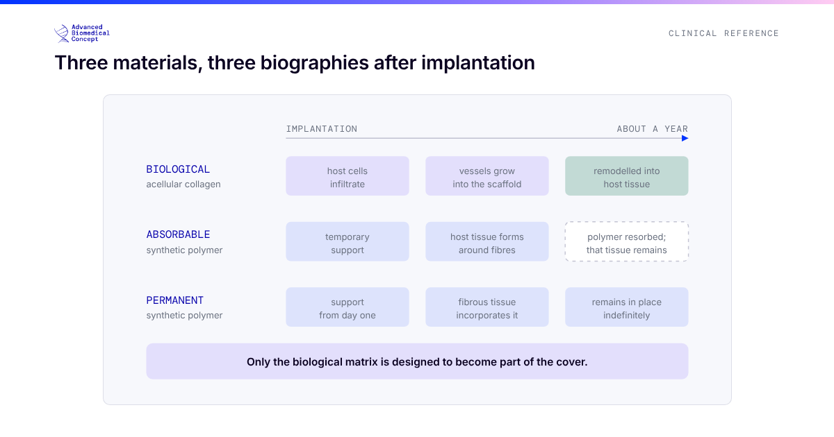
Almost every prepectoral series in the meta-analysis used a matrix or mesh, because prepectorally there is nothing else to define the pocket and hold the implant. That means the prepectoral evidence is, in practice, evidence about prepectoral placement plus a support material, and the choice of material is part of what is being tested.
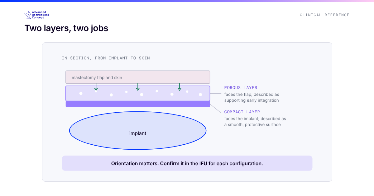
Bovine pericardium has become one of the materials used for this role. Beyond the series above, a multicentre Italian experience with a pericardium pocket in direct-to-implant prepectoral reconstruction reported, across 65 reconstructions and a mean 21 months of follow-up, four minor and two major complications and no significant capsular contracture.6 ExaShape® is a CE-marked bilayer bovine pericardium membrane with pocket configurations designed for this plane, including the BioShield Pocket and its pre-shaped 3D version for a no-touch wrap. The differences between matrix types, and how to read the studies that compare them, are covered in comparing mesh studies and biologic versus synthetic mesh.
What prepectoral reconstruction still asks for
The price of the prepectoral plane is soft-tissue cover. With no muscle over the upper pole, implant edges and rippling show more readily in thin patients. In the pericardium pocket series, 7 of 65 reconstructions had fat grafting at six months for rippling or wrinkling;6 in a series selected specifically for flaps thinner than 1 cm, rippling occurred in 25%.7 Planned staged fat grafting is therefore part of prepectoral reconstruction rather than a complication of it, and the consistency of the graft is what decides how many sessions it takes. Closed-loop processing such as EXAFAT® is designed for that refinement stage.
Cost
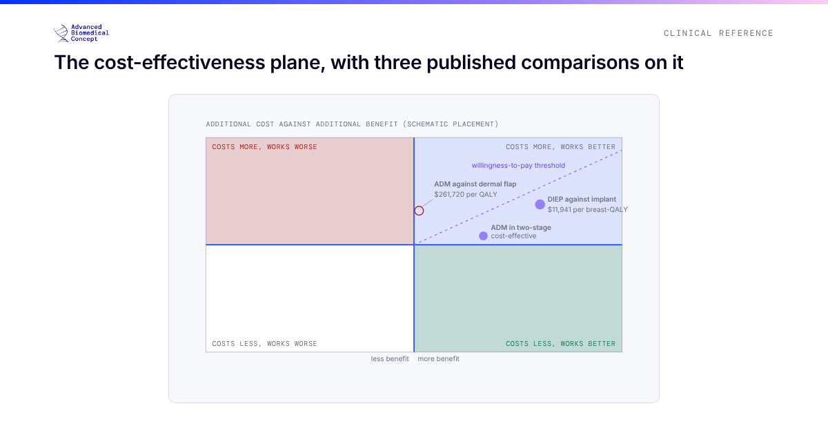
A retrospective analysis of 548 patients at Massachusetts General Hospital found subpectoral reconstruction more costly overall, but the difference disappeared once laterality and staging were accounted for. The authors’ interpretation was that prepectoral reconstruction is cost-neutral in itself and may confer a cost advantage because it is more often performed direct-to-implant.8
The short version
Pooled observational evidence shows prepectoral reconstruction to be at least as safe as subpectoral, with less animation, less capsular contracture and less prosthesis failure, and no outcome in which it does worse. The evidence is consistent but not randomised, and the first randomised trial is under way. Until it reports, the defensible position is the one the data support: prepectoral placement is the better plane for patients whose flaps can carry it, supported by a matrix designed for the job and refined, when needed, with the patient’s own fat.
References
- Potter S, Conroy EJ, Cutress RI, et al. Short-term safety outcomes of mastectomy and immediate implant-based breast reconstruction with and without mesh (iBRA): a multicentre, prospective cohort study. Lancet Oncol. 2019;20(2):254-266. doi:10.1016/S1470-2045(18)30781-2
- Sbitany H, Piper M, Lentz R. Prepectoral breast reconstruction: a safe alternative to submuscular prosthetic reconstruction following nipple-sparing mastectomy. Plast Reconstr Surg. 2017;140(3):432-443. doi:10.1097/PRS.0000000000003627
- Ostapenko E, Nixdorf L, Devyatko Y, Exner R, Wimmer K, Fitzal F. Prepectoral versus subpectoral implant-based breast reconstruction: a systemic review and meta-analysis. Ann Surg Oncol. 2023;30(1):126-136. doi:10.1245/s10434-022-12567-0
- Roberts K, Mills N, Metcalfe C, et al. Best-BRA (Is subpectoral or prepectoral implant placement best in immediate breast reconstruction?): a protocol for a pilot randomised controlled trial. BMJ Open. 2021;11(11):e050886. doi:10.1136/bmjopen-2021-050886
- Nogueira Sixto M, De Castro Parga G, Lamas González MJ, et al. The role of bovine pericardium matrix in immediate prepectoral breast reconstruction: insights from the largest published retrospective series. JPRAS Open. 2025;46:278-289. doi:10.1016/j.jpra.2025.09.005
- De Vita R, Mangialardi ML, Pozzi M, et al. A pericardium bovine matrix pocket in DTI prepectoral breast reconstruction. Clin Breast Cancer. 2024;24(7):e613-e621. doi:10.1016/j.clbc.2024.06.004. Disclosure: the first author is a consultant for ExaShape.
- Casella D, Rocco N, Luridiana G, et al. Enhancing breast reconstruction with bovine pericardium: a preliminary STEP (Surgical Techniques and Efficacy in Pericardium use) towards improved outcomes. J Clin Med. 2025;14(17):6296. doi:10.3390/jcm14176296
- Chinta S, Koh DJ, Sobti N, et al. Cost analysis of pre-pectoral implant-based breast reconstruction. Sci Rep. 2022;12(1):17512. doi:10.1038/s41598-022-21675-6
This article is educational material for healthcare professionals. Plane selection is a judgement about an individual patient; nothing here is a recommendation for a specific case. Series using bovine pericardium matrices other than ExaShape® are cited for the material class, not for the product. ExaShape® and EXAFAT® are CE marked per EU MDR 2017/745 and are for professional use only.
Important information This article is general education, not medical advice. Tap to read the full medical, regulatory & legal notice.
This article is provided by Advanced Biomedical Concept for general informational and educational purposes only. It does not constitute medical, surgical, diagnostic, or other professional healthcare advice, and it is not a substitute for consultation with a suitably qualified healthcare professional. Reading it creates no doctor–patient or other professional relationship. Nothing here should be used to diagnose, treat, cure, or prevent any disease or condition, or to make decisions about medical care. Always seek the advice of a physician or other qualified provider with any questions about a medical condition or treatment, and never disregard or delay professional advice because of something you have read here.
Any reference to Advanced Biomedical Concept products (including ExaShape and ExaFat) is provided for general information only. These are medical devices intended to be used by appropriately trained healthcare professionals strictly in accordance with their applicable, current Instructions for Use (IFU). Regulatory status (including CE marking and classification under the EU Medical Device Regulation), market availability, and approved indications differ between countries and may change over time; a device referred to here may not be available or approved in your country. This content is not intended as, and must not be relied upon as, a statement or claim of safety, clinical performance, or efficacy beyond what is set out in the applicable IFU and current regulatory approvals. Product-specific information is directed at healthcare professionals and is not intended to promote, advertise, or offer any medical device to the general public where such promotion is restricted or prohibited by law.
Statements reflect information believed to be accurate at the date of publication and may be updated or corrected without notice. Any statements regarding the future are expectations or general views, not guarantees. Clinical outcomes depend on many individual factors, results vary from person to person, and no particular result is promised or implied. Where third-party studies, sources, or external links are referenced, they are provided for convenience and information only and do not imply endorsement, and Advanced Biomedical Concept is not responsible for their content.
To the fullest extent permitted by applicable law, Advanced Biomedical Concept and its officers, employees, contributors, and representatives (each acting in that capacity and on behalf of Advanced Biomedical Concept) accept no liability for any loss, injury, or damage of any kind arising directly or indirectly from use of, or reliance on, this content. Nothing in this notice excludes or limits any liability that cannot lawfully be excluded or limited.
Questions about this material can be directed to us via our contact page.
Last reviewed: October 15, 2026.