Where should the implant sit: above the chest muscle, or below it? Few decisions in implant-based breast reconstruction shape the patient’s experience more than this one. Prepectoral reconstruction places the implant above the pectoralis major, leaving the muscle untouched; subpectoral reconstruction places it wholly or partly beneath the muscle. Both are established, both are widely practised, and the right choice depends on the individual patient. This guide compares the two planes on the questions that matter: pain and recovery, animation deformity, complications, radiotherapy and aesthetics, with every figure attributed to its published source.


The two planes, briefly
For decades the subpectoral (or dual-plane) approach was the default. Mastectomy skin flaps were often thin, and lifting the pectoralis major to cover the implant protected it with a layer of vascularised muscle. The cost was collateral: the muscle is detached and stretched over a device it was never meant to cover, which can mean more early pain, functional change and movement of the implant when the muscle contracts.
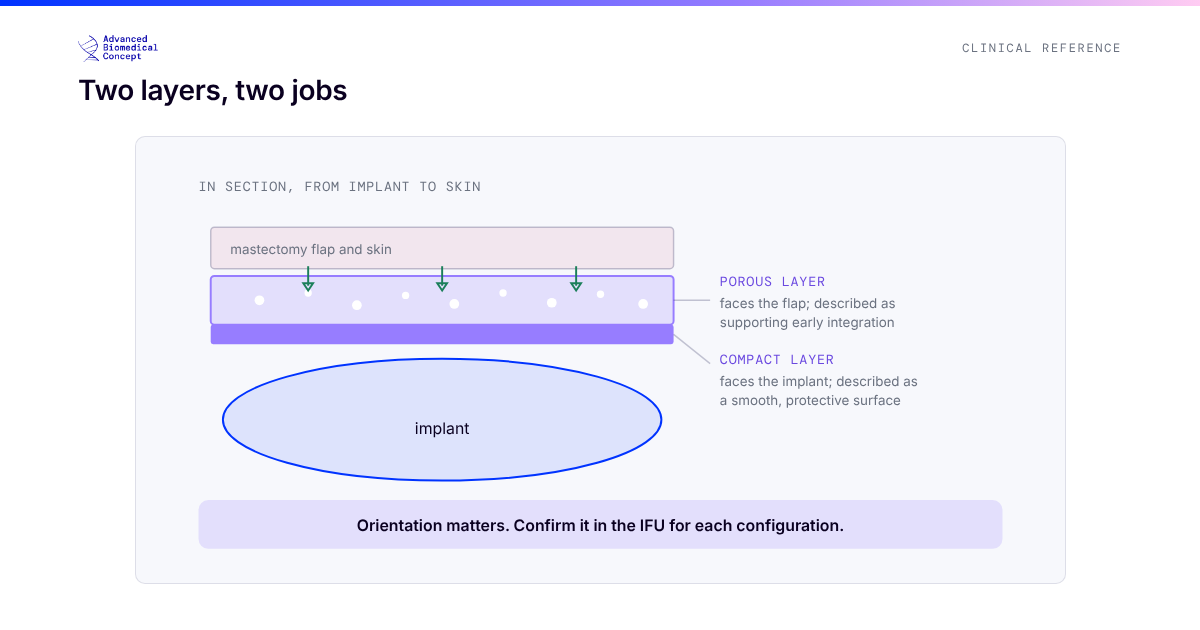
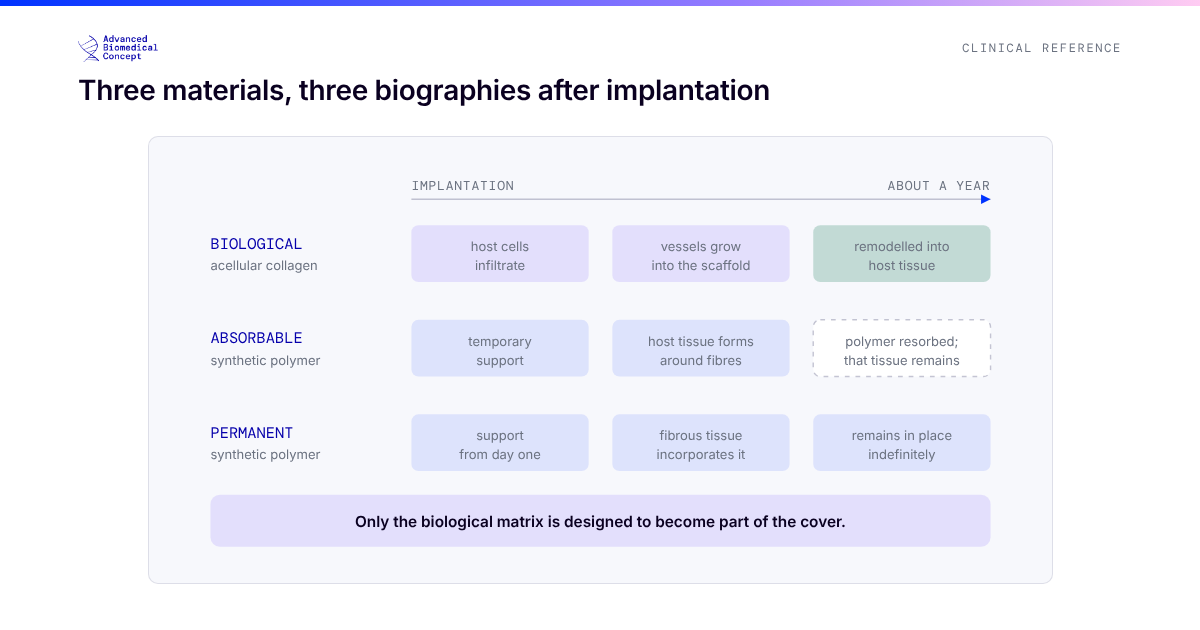
Prepectoral reconstruction became practical when two things matured: better mastectomy technique with careful flap perfusion assessment, and reliable soft-tissue support in the form of a biological matrix or acellular dermal matrix that supports the implant where muscle once did. A joint consensus of UK, European and US breast and plastic surgeons (Highton et al., ecancer 2019) set out the modern framework: with well-perfused flaps and appropriate patient selection, the implant no longer has to go under the muscle.
Pain and recovery
Because the pectoralis major is left in place, prepectoral reconstruction avoids muscle elevation, and several comparative series report less early postoperative pain, lower analgesia requirements and quicker return of upper-limb function. Individual results vary with mastectomy extent, drains and the patient’s baseline, so the honest summary is that sparing the muscle removes one specific source of pain rather than guaranteeing a painless recovery.
Animation deformity
Animation deformity, visible distortion and displacement of the reconstructed breast when the chest muscle contracts, is a phenomenon of submuscular placement; reported rates after subpectoral reconstruction have been substantial in published series, while a prepectoral pocket removes the mechanism entirely, because no muscle passes over the implant. It matters enough that pocket conversion from subpectoral to prepectoral has become a recognised corrective operation (Lentz et al., Gland Surgery 2019). We cover the condition in depth in our article on animation deformity after breast reconstruction.
Complications: what comparative data show
Meta-analyses comparing the planes in immediate implant-based reconstruction generally report broadly comparable overall complication rates, with differences concentrated in specific outcomes rather than across the board. Readers should keep three caveats in mind: cohorts differ in patient selection, matrix use differs between arms, and follow-up lengths vary. Representative findings include:
- Overall complications: systematic reviews of prepectoral versus subpectoral immediate reconstruction report no consistent significant difference in total complications between planes.
- Animation deformity: essentially confined to the subpectoral group, as above.
- Capsular contracture: comparative data are heterogeneous; in one prepectoral bovine pericardium direct-to-implant series (De Vita et al., Clinical Breast Cancer 2024, 65 breasts) no significant capsular contracture was observed at a mean follow-up of 21.3 months, while subpectoral cohorts in the wider literature report low single-digit to double-digit rates depending on radiotherapy and follow-up. These are separate cohorts, not head-to-head comparisons.
- Seroma: some series report more seroma in prepectoral pockets; drain protocols and matrix choice appear to influence this.
For a visual, sourced overview of published outcome figures for the main soft-tissue support options, see our clinical evidence at a glance page.
Radiotherapy
Post-mastectomy radiotherapy (PMRT) raises complication risk in every form of implant-based reconstruction. Comparative work specific to plane, including an Annals of Surgical Oncology analysis of PMRT in prepectoral versus subpectoral reconstruction (Sinnott et al., 2018), suggests prepectoral placement does not worsen, and may reduce, some radiation-associated problems such as capsular contracture severity, though the evidence base is still maturing. We examine this in detail in our dedicated article on radiotherapy and implant-based reconstruction.
Aesthetics: rippling and contour
The trade-off for sparing the muscle is thinner soft-tissue cover over the implant, so visible rippling and palpable edges are the characteristic aesthetic risks of the prepectoral plane, particularly in slim patients. Surgeons manage this with careful patient selection, matrix support and, frequently, staged autologous fat grafting to thicken the envelope; in the De Vita 2024 series, roughly one breast in nine needed fat-graft rippling correction at six months. Subpectoral placement gives more upper-pole padding at the cost of the muscle-related issues above.
How surgeons choose
Modern selection is anatomical and oncological rather than dogmatic. Well-perfused mastectomy flaps, adequate soft-tissue thickness and patient priorities such as avoiding animation and faster recovery favour a prepectoral approach; very thin flaps, compromised perfusion or specific reconstructive goals can favour subpectoral cover. Consensus guidance stresses assessing flap viability at the time of mastectomy and keeping both options open. Our interactive reconstruction options explorer shows where each plane sits among the wider set of choices, and our prepectoral pillar guide covers the muscle-sparing pathway end to end.
The bottom line
Neither plane is universally superior. Subpectoral reconstruction remains a robust, well-understood technique; prepectoral reconstruction spares the muscle, eliminates animation deformity and is supported by a rapidly growing evidence base, at the price of demanding good flaps and thoughtful management of the soft-tissue envelope. The best outcomes come from matching the plane to the patient, which is exactly how the consensus literature frames it.
Key published sources
- Highton L, et al. Prepectoral implant-based breast reconstruction: a joint consensus guide. ecancermedicalscience. 2019.
- De Vita R, et al. Prepectoral direct-to-implant reconstruction with acellular bovine pericardium matrix, multicentre series. Clinical Breast Cancer. 2024.
- Sinnott CJ, et al. Impact of postmastectomy radiation therapy in prepectoral versus subpectoral implant-based breast reconstruction. Annals of Surgical Oncology. 2018.
- Lentz R, et al. Correction of animation deformity with subpectoral to prepectoral implant exchange. Gland Surgery. 2019.
Important information This article is general education, not medical advice. Tap to read the full medical, regulatory & legal notice.
This article is provided by Advanced Biomedical Concept for general informational and educational purposes only. It does not constitute medical, surgical, diagnostic, or other professional healthcare advice, and it is not a substitute for consultation with a suitably qualified healthcare professional. Reading it creates no doctor–patient or other professional relationship. Nothing here should be used to diagnose, treat, cure, or prevent any disease or condition, or to make decisions about medical care. Always seek the advice of a physician or other qualified provider with any questions about a medical condition or treatment, and never disregard or delay professional advice because of something you have read here.
Any reference to Advanced Biomedical Concept products (including ExaShape and ExaFat) is provided for general information only. These are medical devices intended to be used by appropriately trained healthcare professionals strictly in accordance with their applicable, current Instructions for Use (IFU). Regulatory status (including CE marking and classification under the EU Medical Device Regulation), market availability, and approved indications differ between countries and may change over time; a device referred to here may not be available or approved in your country. This content is not intended as, and must not be relied upon as, a statement or claim of safety, clinical performance, or efficacy beyond what is set out in the applicable IFU and current regulatory approvals. Product-specific information is directed at healthcare professionals and is not intended to promote, advertise, or offer any medical device to the general public where such promotion is restricted or prohibited by law.
Statements reflect information believed to be accurate at the date of publication and may be updated or corrected without notice. Any statements regarding the future are expectations or general views, not guarantees. Clinical outcomes depend on many individual factors, results vary from person to person, and no particular result is promised or implied. Where third-party studies, sources, or external links are referenced, they are provided for convenience and information only and do not imply endorsement, and Advanced Biomedical Concept is not responsible for their content.
To the fullest extent permitted by applicable law, Advanced Biomedical Concept and its officers, employees, contributors, and representatives (each acting in that capacity and on behalf of Advanced Biomedical Concept) accept no liability for any loss, injury, or damage of any kind arising directly or indirectly from use of, or reliance on, this content. Nothing in this notice excludes or limits any liability that cannot lawfully be excluded or limited.
Questions about this material can be directed to us via our contact page.
Last reviewed: August 24, 2026.