The move from subpectoral to prepectoral implant placement is the most consequential change in implant-based breast reconstruction of the last decade, and it is frequently described as though one plane simply replaced the other. It did not. The planes trade different problems against each other, and the trade only favours the prepectoral position when the flap can support it.
What the plane actually changes
The choice determines what lies between the implant and the skin, and what the implant is attached to.
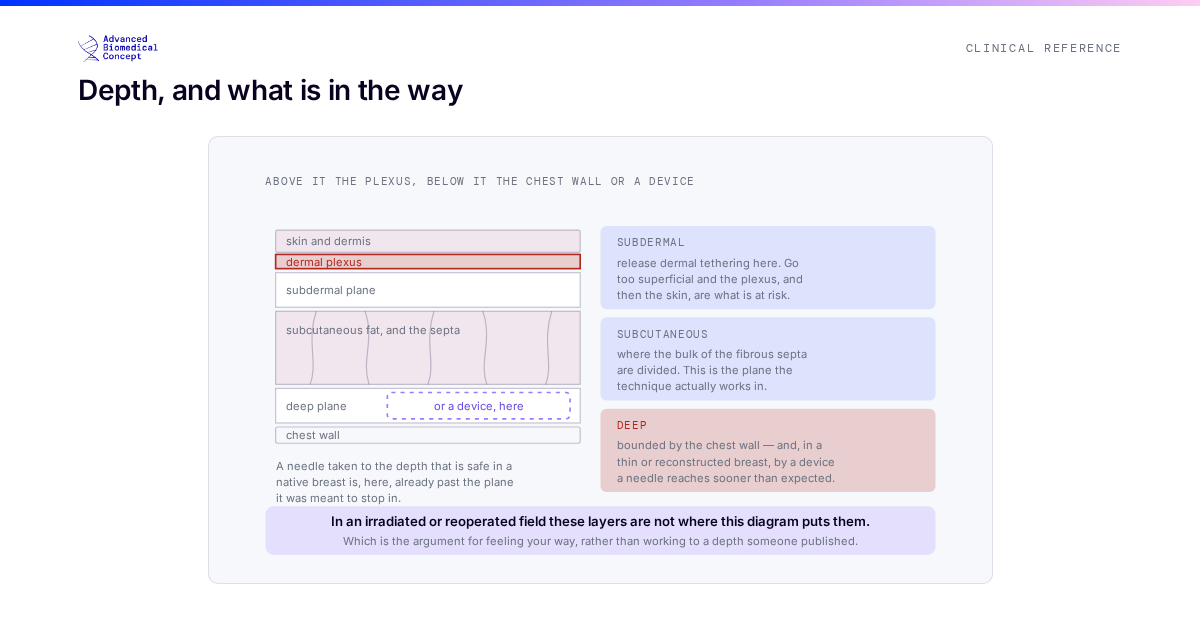
In the subpectoral position, the pectoralis major is elevated and its lower border divided so the muscle can cover the upper portion of the implant. The muscle supplies vascularised tissue between the device and a potentially thin mastectomy flap, and some coverage of the upper pole. The cost is that the muscle has been detached from its insertion and now sits over a device that it contracts across.
In the prepectoral position the muscle is left whole, in its anatomical position, doing its anatomical job. The implant sits in front of it, wrapped or supported by a matrix. Nothing is between the device and the skin except the flap and the matrix — which is why the flap assessment discussed in reading the mastectomy flap stops being one input among several and becomes the deciding one.
The trade, laid out
Animation deformity. A divided, device-covering pectoralis contracts across the implant, producing visible distortion on activation. This is intrinsic to the subpectoral position, not a technical error, and it is a genuine quality-of-life issue for active patients. It is essentially absent prepectorally.
Postoperative pain and recovery. Elevating and dividing muscle hurts more than not doing so, and patients generally report an easier early recovery after prepectoral placement. See recovery after implant reconstruction.
Flap dependence. This is the cost. Prepectorally, every demand for soft-tissue cover falls on the mastectomy flap. A thin or marginally perfused flap that would have been partly protected by muscle is, prepectorally, the only thing between the implant and the outside.
Rippling and contour. With less tissue between device and skin, implant edges and surface irregularity are more visible prepectorally, particularly in the upper pole in thin patients. This is one of the principal roles for fat grafting as a later refinement stage — and the geometry of grafting over an implant under thin skin is exactly the situation described in what determines fat graft retention.
Capsular contracture and radiotherapy. Behaviour in the irradiated field is an area where practice is still consolidating and where the evidence is genuinely mixed; see radiotherapy and breast reconstruction.
The matrix is not optional prepectorally
Subpectoral placement can be done with or without a matrix; the muscle provides some of what the matrix would. Prepectoral placement generally cannot. Something has to control the position of the implant, define the lower pole, and distribute load across the pocket rather than concentrating it on the flap, and in the prepectoral position that is the matrix’s job.
Which means matrix selection stops being a preference and becomes part of the reconstruction’s structural design: how it integrates, how quickly it revascularises, how it behaves under load and how it handles. Those properties are compared in types of biological matrices, xenograft versus allograft and matrix versus ADM versus synthetic mesh. Integration specifically — the process by which the matrix stops being an implanted sheet and becomes the patient’s own tissue — is covered in how a biological matrix integrates.
Selection, in practice
Reduced to its essentials, the question is whether the flap can carry the reconstruction alone. Where it can, prepectoral placement avoids a set of muscle-related problems at the cost of a set of coverage-related ones that are manageable and largely correctable. Where it cannot, the muscle is doing something useful and removing it in pursuit of a better recovery profile is a poor trade.
Ptosis, breast width, native tissue thickness, the planned incision, whether radiotherapy is expected, and the patient’s own priorities all inform the choice. A competitive swimmer and a sedentary patient weight animation deformity very differently, and that is a legitimate input rather than a soft one — which is the argument for collecting patient-reported outcomes systematically, as discussed in patient-reported outcomes and BREAST-Q.
Changing plane later is not symmetrical
Plane selection is often discussed as though it could be revisited at revision. It can, but not equally in both directions, and the asymmetry is worth knowing before the first operation.
Converting subpectoral to prepectoral — “site change” — is a recognised revision for animation deformity and is done routinely. The muscle is returned to the chest wall, the implant moves in front of it, and a matrix takes over the support role. It works because the muscle, once released from its device-covering position, has somewhere to go back to.
The reverse is harder. A patient reconstructed prepectorally whose flap subsequently thins, or who develops rippling that fat grafting cannot correct, cannot simply be moved behind a muscle that was never elevated and that now sits over a mature capsule. It is not impossible, but it is a more substantial operation than the conversion in the other direction.
The practical implication is that in a genuinely borderline case, the subpectoral position preserves more future options. That is not an argument for defaulting to it — the animation and recovery costs are real and are paid by every patient, not just the borderline ones — but it belongs in the reasoning.
Reading the comparative evidence
Comparisons between the planes are complicated by the fact that the prepectoral cohorts are, by design, the patients with better flaps. Selection runs in the direction of the result, and a series showing lower complication rates prepectorally may be reporting the quality of the selection rather than the superiority of the plane. The questions worth asking of such a paper are in reading a breast reconstruction clinical study, and overall complication figures are discussed in complication rates in implant reconstruction.
The short version
Prepectoral placement moves the burden from the muscle to the flap. That is an improvement when the flap can carry it and a mistake when it cannot, and the assessment that decides which is being made in theatre, on the day, by the surgeon looking at the tissue.
This article is educational material for clinicians. It sets out a trade-off rather than a recommendation; plane selection is a judgement about an individual patient, their tissue and their priorities.
Important information This article is general education, not medical advice. Tap to read the full medical, regulatory & legal notice.
This article is provided by Advanced Biomedical Concept for general informational and educational purposes only. It does not constitute medical, surgical, diagnostic, or other professional healthcare advice, and it is not a substitute for consultation with a suitably qualified healthcare professional. Reading it creates no doctor–patient or other professional relationship. Nothing here should be used to diagnose, treat, cure, or prevent any disease or condition, or to make decisions about medical care. Always seek the advice of a physician or other qualified provider with any questions about a medical condition or treatment, and never disregard or delay professional advice because of something you have read here.
Any reference to Advanced Biomedical Concept products (including ExaShape and ExaFat) is provided for general information only. These are medical devices intended to be used by appropriately trained healthcare professionals strictly in accordance with their applicable, current Instructions for Use (IFU). Regulatory status (including CE marking and classification under the EU Medical Device Regulation), market availability, and approved indications differ between countries and may change over time; a device referred to here may not be available or approved in your country. This content is not intended as, and must not be relied upon as, a statement or claim of safety, clinical performance, or efficacy beyond what is set out in the applicable IFU and current regulatory approvals. Product-specific information is directed at healthcare professionals and is not intended to promote, advertise, or offer any medical device to the general public where such promotion is restricted or prohibited by law.
Statements reflect information believed to be accurate at the date of publication and may be updated or corrected without notice. Any statements regarding the future are expectations or general views, not guarantees. Clinical outcomes depend on many individual factors, results vary from person to person, and no particular result is promised or implied. Where third-party studies, sources, or external links are referenced, they are provided for convenience and information only and do not imply endorsement, and Advanced Biomedical Concept is not responsible for their content.
To the fullest extent permitted by applicable law, Advanced Biomedical Concept and its officers, employees, contributors, and representatives (each acting in that capacity and on behalf of Advanced Biomedical Concept) accept no liability for any loss, injury, or damage of any kind arising directly or indirectly from use of, or reliance on, this content. Nothing in this notice excludes or limits any liability that cannot lawfully be excluded or limited.
Questions about this material can be directed to us via our contact page.
Last reviewed: September 25, 2026.