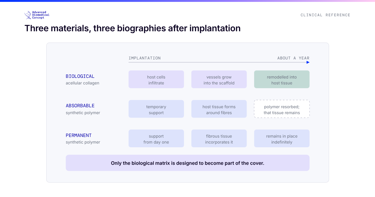
“Mesh” in breast reconstruction covers a family of materials that do not have much in common beyond their job. Human dermis, porcine and bovine dermis, bovine pericardium, absorbable synthetic knits and permanent titanised polymers are all placed around implants for broadly the same reason, and they behave very differently once they are there. The literature on them is large, uneven, and frequently summarised badly, because studies of one material are cited as if they described all of them.
This article sets out what meshes and matrices are for, how the families differ, what the best evidence says about using one at all, and how the question changes in the prepectoral plane, where it stops being optional.
What a mesh is for
In implant reconstruction, a support material does up to four jobs. It extends cover where native tissue is insufficient, classically the lower pole beneath a pectoralis muscle that has been released. It defines the pocket, controlling where the implant sits and stopping it migrating laterally or inferiorly. It sets the inframammary fold. And, in the prepectoral plane, it takes the place of the muscle entirely, standing between the implant and the mastectomy flap.
The material families
Every support material in breast reconstruction belongs to one of two groups, and the biological group divides further by source tissue.
Bovine pericardium sits apart within the biological group. It is a thinner, naturally layered collagen membrane with a lower biological mass per unit area than dermis. In a rat model of breast implant coverage, acellular pericardial meshes reduced capsular α-SMA expression and inflammatory cell infiltration and increased capillary density, and the structure of the individual product influenced how the tissue remodelled.1 That is preclinical evidence and should be held at that level, but it describes the kind of difference that makes pooling “biological meshes” into one category misleading. Material processing and integration are covered in more depth in how a biological matrix integrates.
What the evidence says about using one at all
The national picture. The UK iBRA study captured 2,108 women having immediate implant reconstruction at 81 units between 2014 and 2016. Two-thirds had a mesh: 54% biological, 12% synthetic. At three months, 9% had lost their implant, 18% had been readmitted, 18% had returned to theatre and 25% had needed treatment for infection, all above the national quality standards.2 iBRA was not designed to compare mesh with no mesh, but it set the baseline: implant reconstruction as practised at scale had a higher complication burden than surgeons assumed, and much of it involved mesh.
The randomised trials. A Swedish and UK trial randomised 135 women to subpectoral reconstruction with or without a porcine dermal matrix. At six months, implant loss was the same in both arms (6%), but the matrix arm showed a trend towards more complications and reoperations, and significantly more wound-healing problems.3 The Dutch BRIOS trial, discussed in detail in DTI versus two-stage, found one-stage reconstruction with the same type of matrix markedly worse than two-stage reconstruction.4 Both trials tested porcine dermis in the subpectoral plane.
The network meta-analysis. The most useful synthesis so far is a network meta-analysis of 31 studies by Murphy and colleagues, which compared human dermis, xenogeneic dermis and synthetic mesh with traditional submuscular reconstruction without any support. Human dermis was associated with significantly more overall complications, infection and seroma than no mesh. Xenogeneic matrices and synthetic mesh were not significantly different from no mesh for overall complications. No option was superior for implant loss.5
Two conclusions follow. The first is that the material matters: the excess complications attributed to “ADM” in much of the literature are concentrated in one family. The second is that all of this is subpectoral evidence. When the comparator is a muscle pocket that can, in principle, do without support, the question is whether a mesh adds enough to justify itself. In the prepectoral plane there is no such comparator.
In the prepectoral plane, the question changes
Prepectorally, something has to hold the implant. The relevant question is no longer whether to use a support material but which one, configured how, and in which patient. That makes the properties of the material part of the design of the reconstruction: how quickly it integrates and revascularises, how much biological mass it adds under a thin flap, whether it encourages fluid collection, how it handles, and whether it can be shaped to the implant before insertion.
Technique matters as much as material. A series from Sassari illustrates this with a dermal matrix. Changing how the matrix was fixed to the chest wall in prepectoral direct-to-implant reconstruction was associated with a reduction in major complications, including seroma from 13.9% to 2.3% and implant loss from 7% to 0%.6 This is a retrospective before-and-after comparison, but it shows how much of the outcome sits in configuration rather than in the label on the box.
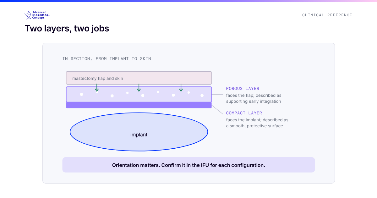
That is the logic behind pre-shaped devices. ExaShape® is a CE-marked bilayer bovine pericardium membrane, with a compact layer facing the implant and a porous layer facing the flap, supplied in configurations designed for specific jobs: the BioShield Pocket and 3D Pocket wrap the anterior implant for prepectoral reconstruction, PREPEC covers selected parts of the implant, Grid supports the lower pole subpectorally, and dedicated versions exist for tissue expanders and the nipple-areola complex. The multicentre experience with the pericardium pocket in direct-to-implant prepectoral reconstruction, 65 reconstructions over a mean 21 months, reported four minor and two major complications.7 Practical selection between configurations is covered in the practical ExaShape guide.
Cost is part of the evidence
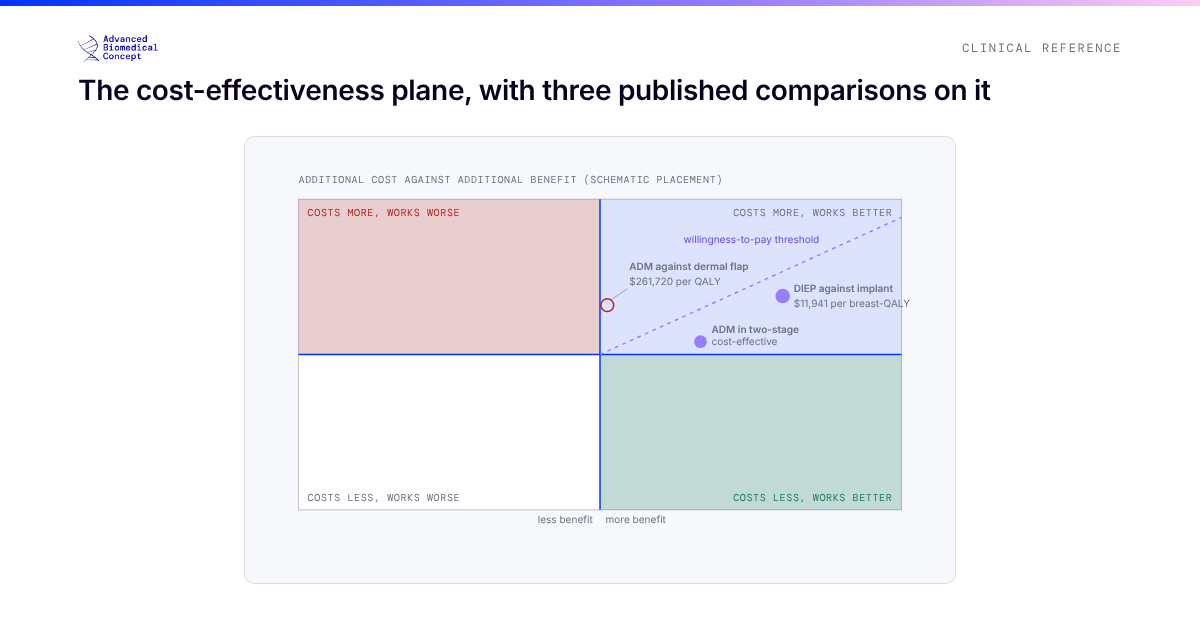
Support materials are expensive, and whether they are worth it depends on what they prevent. A decision model of two-stage expander reconstruction found a human dermal matrix cost-effective, adding $361.96 per case for an estimated gain in quality-adjusted life years.8 The same group found the same material not cost-effective in single-stage reconstruction when an autologous dermal flap was a feasible alternative.9 Context decides. The economics are discussed in full in the cost-effectiveness of reconstruction.
The short version
Meshes are a family, not a product. In the subpectoral plane the evidence for adding one is mixed and depends heavily on the material, with the clearest excess of complications in human dermis. In the prepectoral plane a support material is structural, so the evidence that matters is about which material, in which configuration, placed how. That is where the field’s attention, and the case for purpose-designed devices, now sits.
References
- Bernardini R, Varvaras D, D’Amico F, et al. Biological acellular pericardial mesh regulated tissue integration and remodeling in a rat model of breast prosthetic implantation. J Biomed Mater Res B Appl Biomater. 2020;108(2):577-590. doi:10.1002/jbm.b.34413
- Potter S, Conroy EJ, Cutress RI, et al. Short-term safety outcomes of mastectomy and immediate implant-based breast reconstruction with and without mesh (iBRA): a multicentre, prospective cohort study. Lancet Oncol. 2019;20(2):254-266. doi:10.1016/S1470-2045(18)30781-2
- Lohmander F, Lagergren J, Roy PG, et al. Implant based breast reconstruction with acellular dermal matrix: safety data from an open-label, multicenter, randomized, controlled trial in the setting of breast cancer treatment. Ann Surg. 2019;269(5):836-841. doi:10.1097/SLA.0000000000003054
- Dikmans REG, Negenborn VL, Bouman MB, et al. Two-stage implant-based breast reconstruction compared with immediate one-stage implant-based breast reconstruction augmented with an acellular dermal matrix: an open-label, phase 4, multicentre, randomised, controlled trial. Lancet Oncol. 2017;18(2):251-258. doi:10.1016/S1470-2045(16)30668-4
- Murphy D, O’Donnell JP, Ryan ÉJ, et al. Immediate breast cancer reconstruction with or without dermal matrix or synthetic mesh support: a review and network meta-analysis. Plast Reconstr Surg. 2023;151(4):563e-574e. doi:10.1097/PRS.0000000000009984
- Tettamanzi M, Arrica G, Ziani F, et al. Prepectoral breast reconstruction with prosthesis and acellular dermal matrix: a new technique of ADM implantation and fixation. Plast Reconstr Surg Glob Open. 2024;12(6):e5895. doi:10.1097/GOX.0000000000005895
- De Vita R, Mangialardi ML, Pozzi M, et al. A pericardium bovine matrix pocket in DTI prepectoral breast reconstruction. Clin Breast Cancer. 2024;24(7):e613-e621. doi:10.1016/j.clbc.2024.06.004. Disclosure: the first author is a consultant for ExaShape.
- Krishnan NM, Chatterjee A, Rosenkranz KM, et al. The cost effectiveness of acellular dermal matrix in expander-implant immediate breast reconstruction. J Plast Reconstr Aesthet Surg. 2014;67(4):468-476. doi:10.1016/j.bjps.2013.12.035
- Krishnan NM, Chatterjee A, Van Vliet MM, et al. A comparison of acellular dermal matrix to autologous dermal flaps in single-stage, implant-based immediate breast reconstruction: a cost-effectiveness analysis. Plast Reconstr Surg. 2013;131(5):953-961. doi:10.1097/PRS.0b013e3182865a24
This article is educational material for healthcare professionals. It describes published evidence on classes of support material and does not claim clinical superiority for any product. ExaShape® is CE marked per EU MDR 2017/745 and is for professional use only.
Important information This article is general education, not medical advice. Tap to read the full medical, regulatory & legal notice.
This article is provided by Advanced Biomedical Concept for general informational and educational purposes only. It does not constitute medical, surgical, diagnostic, or other professional healthcare advice, and it is not a substitute for consultation with a suitably qualified healthcare professional. Reading it creates no doctor–patient or other professional relationship. Nothing here should be used to diagnose, treat, cure, or prevent any disease or condition, or to make decisions about medical care. Always seek the advice of a physician or other qualified provider with any questions about a medical condition or treatment, and never disregard or delay professional advice because of something you have read here.
Any reference to Advanced Biomedical Concept products (including ExaShape and ExaFat) is provided for general information only. These are medical devices intended to be used by appropriately trained healthcare professionals strictly in accordance with their applicable, current Instructions for Use (IFU). Regulatory status (including CE marking and classification under the EU Medical Device Regulation), market availability, and approved indications differ between countries and may change over time; a device referred to here may not be available or approved in your country. This content is not intended as, and must not be relied upon as, a statement or claim of safety, clinical performance, or efficacy beyond what is set out in the applicable IFU and current regulatory approvals. Product-specific information is directed at healthcare professionals and is not intended to promote, advertise, or offer any medical device to the general public where such promotion is restricted or prohibited by law.
Statements reflect information believed to be accurate at the date of publication and may be updated or corrected without notice. Any statements regarding the future are expectations or general views, not guarantees. Clinical outcomes depend on many individual factors, results vary from person to person, and no particular result is promised or implied. Where third-party studies, sources, or external links are referenced, they are provided for convenience and information only and do not imply endorsement, and Advanced Biomedical Concept is not responsible for their content.
To the fullest extent permitted by applicable law, Advanced Biomedical Concept and its officers, employees, contributors, and representatives (each acting in that capacity and on behalf of Advanced Biomedical Concept) accept no liability for any loss, injury, or damage of any kind arising directly or indirectly from use of, or reliance on, this content. Nothing in this notice excludes or limits any liability that cannot lawfully be excluded or limited.
Questions about this material can be directed to us via our contact page.
Last reviewed: October 15, 2026.