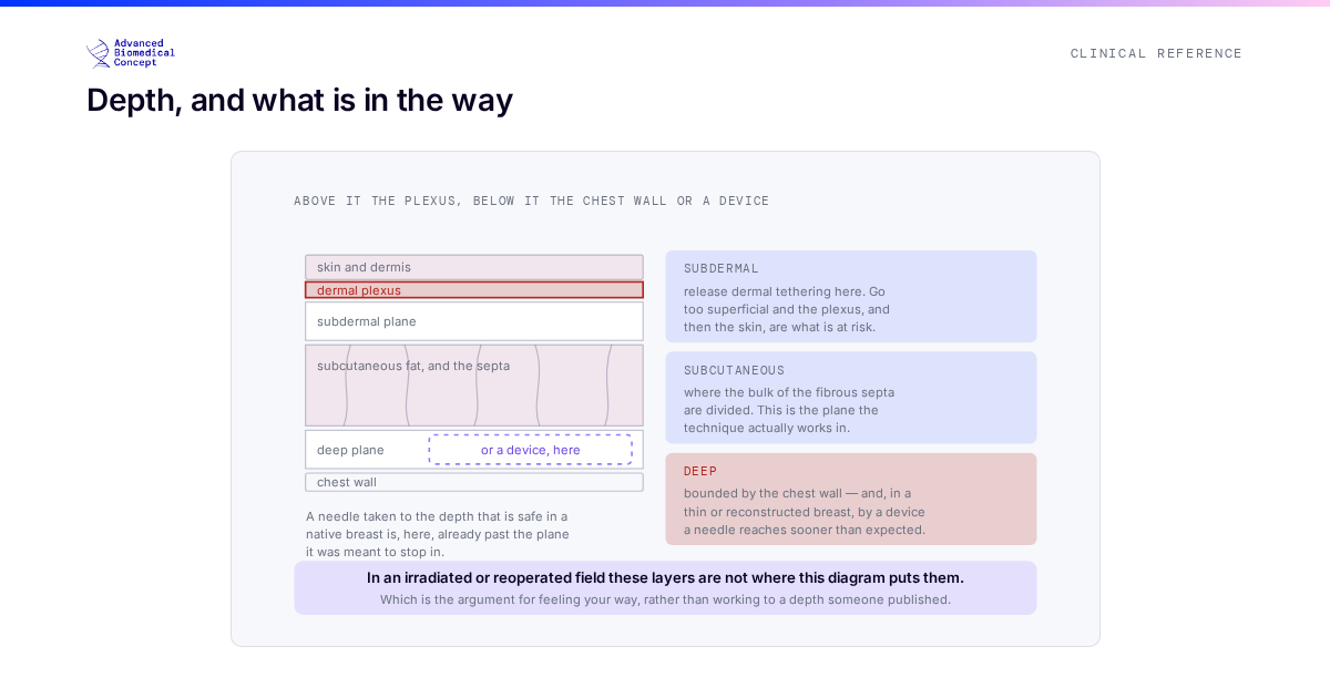
Percutaneous release is a deceptively simple manoeuvre with one difficult constraint: the releases must not join up. Everything that distinguishes a well-executed rigottomy from a counterproductive one follows from that.
This article is about the mechanics — what the release is doing to the tissue, why the pattern matters more than the number of passes, and how the commonest technical error produces precisely the complication the technique exists to prevent.
What is being released
A fibrous or scarred recipient bed is not simply dense; it is tethered. Fibrous septa and a contracted aponeurotic layer hold the tissue as a plate, resisting expansion in every direction and leaving no interstitial space for a graft to occupy. Fat injected into such a plate has nowhere to distribute to. It follows the path of least resistance, which is usually a single plane, and collects.
Release divides those bands through needle-sized skin punctures, without an open incision. The tissue that was a plate becomes a lattice.
Two features of the third panel carry the whole technique. The cavities are numerous, so there is a large internal surface area in contact with released, vascularised tissue. And they are separate, so each remains an individual space of a size the graft can survive in.
The error that turns the technique against itself
If releases at the same depth run into one another, the individual cavities coalesce. The result is not a bigger lattice. It is one large cavity — a macrocavity — and a macrocavity is the exact geometry that fat grafting is trying to avoid.
This is worth stating carefully, because it is counterintuitive. The fat is identical. The volume is identical. The bed is not worse in any way that could be measured before the graft was placed. But in the second case the graft cannot organise itself into parcels that diffusion can support, because the space it was given has no internal walls. It pools, the rim takes, the centre does not, and the sequence runs on: fat necrosis, oil cyst, palpable lump, a patient returning for imaging and often biopsy, and a lesion that has to be distinguished from recurrence.
The surgeon did not place the fat badly. The surgeon made the space badly, several minutes earlier.
What separates the cavities, in practice
Three things, and they reinforce each other.
Varying the depth. Releases made at staggered depths cannot coalesce even where they cross in plan view. This is the most reliable of the three because it does not depend on precise spacing.
Varying the angle. Passes made along a single axis divide the same bands repeatedly and tend to open a plane. Passes made across several axes divide different bands and leave the tissue between them intact.
Restraint in extent. Each individual release is small — the manoeuvre is a division of a band, not a dissection. A pass that travels is a pass that has connected two cavities.
The counterintuitive summary is that a more aggressive release is not a better one. The endpoint is compliance with the internal structure preserved, and a bed that has been over-released has traded its structure for its compliance.
Knowing when it is done
The endpoint is tactile. The tissue lifts and spreads where it previously would not; the skin redrapes; tethering on the surface releases. It is a change in behaviour rather than a count of passes, which is why the number of releases is a poor way to describe or teach the technique and why the assessment has to be made continuously rather than at the end.
Over-release announces itself too, as a bed that feels loose rather than compliant — and by then the internal walls are gone.
The technique is performed blind, and that has consequences
Everything above describes an architecture built without seeing it. There is no direct vision; the surgeon knows where the instrument is by feel, by external landmarks, and by what the tissue does in response. Two consequences follow, and both are underappreciated.
The first is bleeding. Dividing fibrous bands divides small vessels running with them, and the resulting bleeding is into the cavities that were just created. Blood occupying a micro-cavity is blood displacing the graft, and a haematoma in a released bed is both a space-occupying problem and an infection risk. This is part of the argument for a release that is adequate rather than maximal: more division means more divided vessels.
The second is that the result cannot be inspected. There is no moment at which the surgeon sees the lattice and confirms it looks as intended. The endpoint is inferred from tissue behaviour, which means the technique is unusually dependent on the operator having a clear mental model of what they are constructing — and unusually unforgiving of one who is simply making passes until the tissue gives.
It is also why the complication presents so late and so misleadingly. A macrocavity created in theatre produces no sign at the time, no sign at the first dressing change, and becomes apparent as a lump months later, at which point it is naturally attributed to the graft.
Release and graft are one operation
The two steps are not independent. Release creates a specific internal architecture, and the graft is placed to populate it — small parcels into many cavities, not a volume into a pocket. A carefully created lattice filled by a single large deposit has wasted the release entirely, and this is why the same surgeon performs both and why the grafting technique has to match the release pattern that was actually made.
The wider role of release as bed preparation is covered in rigottomy as recipient-site preparation, the underlying principle in what rigottomy is, and the reason the geometry matters at all in what determines fat graft retention.
Where it is used
The technique originated in scar and burn contracture and remains a mainstay there. In breast surgery it appears in the constricting ring of a tuberous breast, described in rigottomy for tuberous breast and scar release; in the tethered and thin chest wall after mastectomy; and in the irradiated field, where both the release and its timing require care — the subject of depth, planes and radiotherapy timing.
What patients are told about recovery and staging is set out in is rigottomy safe: recovery and what to expect.
The short version
The technique is not “cutting the scar”. It is building an internal structure of many separate spaces, each small enough for a graft to survive in. Releases that join up destroy that structure, and the complication that follows is indistinguishable from bad grafting — which is why it is usually blamed on it.
This article is educational material for clinicians. It describes mechanics and failure modes, not a protocol, and gives no depths, spacings or pass counts: those are judgements about the tissue under the hand.
Important information This article is general education, not medical advice. Tap to read the full medical, regulatory & legal notice.
This article is provided by Advanced Biomedical Concept for general informational and educational purposes only. It does not constitute medical, surgical, diagnostic, or other professional healthcare advice, and it is not a substitute for consultation with a suitably qualified healthcare professional. Reading it creates no doctor–patient or other professional relationship. Nothing here should be used to diagnose, treat, cure, or prevent any disease or condition, or to make decisions about medical care. Always seek the advice of a physician or other qualified provider with any questions about a medical condition or treatment, and never disregard or delay professional advice because of something you have read here.
Any reference to Advanced Biomedical Concept products (including ExaShape and ExaFat) is provided for general information only. These are medical devices intended to be used by appropriately trained healthcare professionals strictly in accordance with their applicable, current Instructions for Use (IFU). Regulatory status (including CE marking and classification under the EU Medical Device Regulation), market availability, and approved indications differ between countries and may change over time; a device referred to here may not be available or approved in your country. This content is not intended as, and must not be relied upon as, a statement or claim of safety, clinical performance, or efficacy beyond what is set out in the applicable IFU and current regulatory approvals. Product-specific information is directed at healthcare professionals and is not intended to promote, advertise, or offer any medical device to the general public where such promotion is restricted or prohibited by law.
Statements reflect information believed to be accurate at the date of publication and may be updated or corrected without notice. Any statements regarding the future are expectations or general views, not guarantees. Clinical outcomes depend on many individual factors, results vary from person to person, and no particular result is promised or implied. Where third-party studies, sources, or external links are referenced, they are provided for convenience and information only and do not imply endorsement, and Advanced Biomedical Concept is not responsible for their content.
To the fullest extent permitted by applicable law, Advanced Biomedical Concept and its officers, employees, contributors, and representatives (each acting in that capacity and on behalf of Advanced Biomedical Concept) accept no liability for any loss, injury, or damage of any kind arising directly or indirectly from use of, or reliance on, this content. Nothing in this notice excludes or limits any liability that cannot lawfully be excluded or limited.
Questions about this material can be directed to us via our contact page.
Last reviewed: September 25, 2026.