One surface protects the implant. The other invites the body in. ExaShape does not ask you to choose.
Ask a biological matrix to do two opposite things at once and it usually disappoints at both. It should shield the implant from friction and the surrounding environment, yet it should also welcome vessels, cells, and new collagen. A single, uniform sheet cannot be optimised for a barrier and for integration at the same time. ExaShape resolves that tension by not being uniform at all.
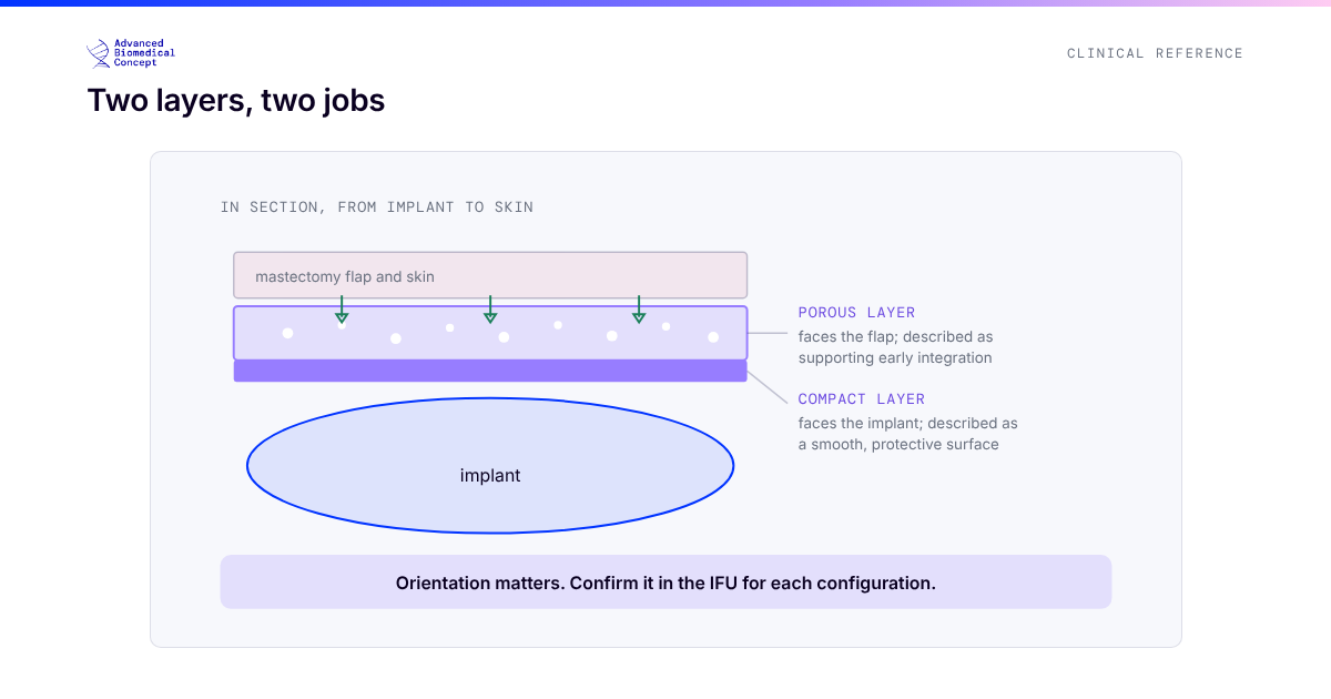
Because it preserves the native structure of bovine pericardium, ExaShape is a true bilayer. Each face has a job.
The compact layer: the implant’s side
The compact layer sits against the device. Its surface is smooth, which reduces friction against the implant shell. It protects the implant from external factors and lends the construct structural support. This is the face that keeps the mechanical relationship between matrix and implant clean and stable.
The porous layer: the body’s side
The porous layer faces the tissue, and its open architecture is where integration begins. That porosity allows an early start to the biological process: progressive neovascularization, fibroblast colonization, and collagen formation. The same open structure supports tissue adherence and implant stability over time. Where the compact layer manages the mechanical interface, the porous layer manages the biological one.
Two layers, four stages
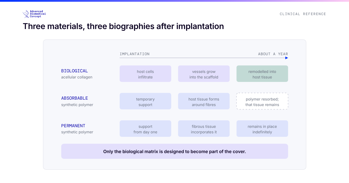
The bilayer is not a static feature. It choreographs a sequence that unfolds over months. ExaShape’s mode of action moves through four stages.
1. Early inflammation. Immediately after implantation, the response is dominated by M2 macrophages associated with tissue repair, and growth factors are released to activate fibroblasts. Because ExaShape carries a low biological mass, this inflammatory response stays minimal, which is associated with a reduced risk of seroma and oedema.
2. Neoangiogenesis. New blood vessels grow inside the porous layer and progressively into the compact layer, driving rapid fibroblast repopulation. The permeability of the porous layer lets this begin earlier, which is associated with a reduced risk of necrosis.
3. Collagen formation. Fibroblasts lay down patient-specific collagen, first filling the porous layer, then the compact layer. ExaShape becomes firmly integrated with the surrounding tissue, turning flexible and biologically active as native collagen replaces the original matrix.
4. Remodelling. Collagenases degrade the original matrix, creating spaces that are progressively replaced by organised collagen and new blood vessels. The regenerated tissue is elastic, biologically active, and thicker than the original membrane.
Why the architecture pays off
The two-layer design is the reason the mode of action reads the way it does. A barrier surface keeps the implant relationship stable while an integration surface pulls the biology forward on schedule. Real-world clinical data reflects this: progressive neovascularization within the matrix has been observed across a 12-month period (Varvaras D, et al. St. Gallen 2023), alongside a regulated inflammatory response and controlled periprosthetic remodelling (Bernardini R, et al. J Biomed Mater Res. 2020).
A matrix that starts as a device and finishes as living, patient-specific tissue is doing something a uniform sheet cannot. That is the point of the bilayer.
One material. Two surfaces. A single, predictable arc from implantation to integration.
See the four-stage mode of action in motion on the ExaShape line page, or request a demo.
Scientific references
- Varvaras D, et al. Use of Acellular Pericardial Biological Mesh for Prepectoral and Dual Plane. St. Gallen 2023.
- Bernardini R, et al. J Biomed Mater Res. 2020.
- De Vita R, et al. A Pericardium Bovine Matrix Pocket in DTI Prepectoral Breast Reconstruction. Clinical Breast Cancer. 2024.
Images for illustrative purposes only. For Healthcare Professionals Only. ExaShape is CE marked per EU MDR 2017/745.
Important information This article is general education, not medical advice. Tap to read the full medical, regulatory & legal notice.
This article is provided by Advanced Biomedical Concept for general informational and educational purposes only. It does not constitute medical, surgical, diagnostic, or other professional healthcare advice, and it is not a substitute for consultation with a suitably qualified healthcare professional. Reading it creates no doctor–patient or other professional relationship. Nothing here should be used to diagnose, treat, cure, or prevent any disease or condition, or to make decisions about medical care. Always seek the advice of a physician or other qualified provider with any questions about a medical condition or treatment, and never disregard or delay professional advice because of something you have read here.
Any reference to Advanced Biomedical Concept products (including ExaShape and ExaFat) is provided for general information only. These are medical devices intended to be used by appropriately trained healthcare professionals strictly in accordance with their applicable, current Instructions for Use (IFU). Regulatory status (including CE marking and classification under the EU Medical Device Regulation), market availability, and approved indications differ between countries and may change over time; a device referred to here may not be available or approved in your country. This content is not intended as, and must not be relied upon as, a statement or claim of safety, clinical performance, or efficacy beyond what is set out in the applicable IFU and current regulatory approvals. Product-specific information is directed at healthcare professionals and is not intended to promote, advertise, or offer any medical device to the general public where such promotion is restricted or prohibited by law.
Statements reflect information believed to be accurate at the date of publication and may be updated or corrected without notice. Any statements regarding the future are expectations or general views, not guarantees. Clinical outcomes depend on many individual factors, results vary from person to person, and no particular result is promised or implied. Where third-party studies, sources, or external links are referenced, they are provided for convenience and information only and do not imply endorsement, and Advanced Biomedical Concept is not responsible for their content.
To the fullest extent permitted by applicable law, Advanced Biomedical Concept and its officers, employees, contributors, and representatives (each acting in that capacity and on behalf of Advanced Biomedical Concept) accept no liability for any loss, injury, or damage of any kind arising directly or indirectly from use of, or reliance on, this content. Nothing in this notice excludes or limits any liability that cannot lawfully be excluded or limited.
Questions about this material can be directed to us via our contact page.
Last reviewed: July 1, 2026.