Advanced Biomedical Concept
Request a Demo For Surgeons For Distributors
🇬🇧 English EN 🇮🇹 Italiano IT 🇫🇷 Français FR 🇩🇪 Deutsch DE 🇪🇸 Español ES 🇵🇹 Português PT 🇹🇷 Türkçe TR 🇸🇦 العربية AR 🇯🇵 日本語 JA

MENU

Request a Demo For Surgeons For Distributors
Category

Clinical Insights

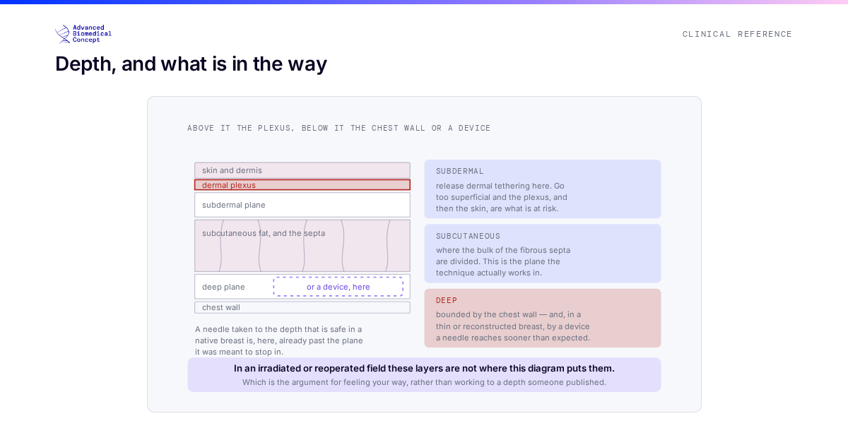
A cross-section through skin, subdermal plane, subcutaneous fat and the deeper layer beneath, with what a release is doing at each depth and what it must not reach. The subdermal plane is where dermal tethering is released and where going too superficial risks the dermal plexus and the skin itself. The subcutaneous plane is where the bulk of fibrous septa are divided. The deep plane is bounded by the chest wall, and in a reconstructed or thin breast by an implant or expander, either of which a needle can reach in tissue far thinner than it looks. A note records that in an irradiated or previously operated field the planes are not where the anatomy says they are.
Clinical Insights — 16 September 2026

Rigottomy in the Irradiated and Reconstructed Chest: Depth and Timing

Two questions decide whether percutaneous release is safe in an irradiated or reconstructed chest: how deep, and when. Neither has a number…

Read article →
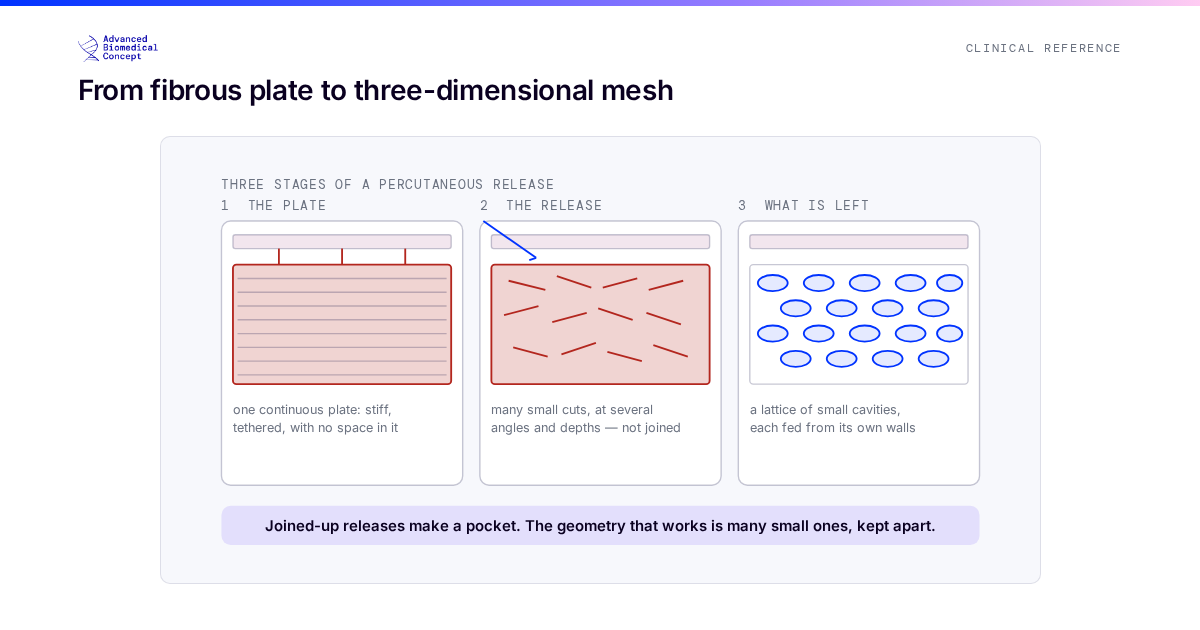
Three stages of a percutaneous release. First, scarred tissue as a continuous fibrous plate under the skin, stiff and tethered, with no space in it. Second, a fine needle passed through the skin at shallow angles making many small independent cuts through the fibrous bands, in several directions and at several depths, without joining them up. Third, the result: a lattice of small separate cavities held open by the tension that was previously locked in the plate, each cavity small enough to be fed from its own walls. The figure marks that the releases are deliberately not connected to one another.
Clinical Insights — 13 September 2026

Rigottomy Release Mechanics: Why the Cuts Must Not Join Up

The technique is not cutting the scar; it is building many separate spaces small enough for a graft to survive in. Releases…

Read article →
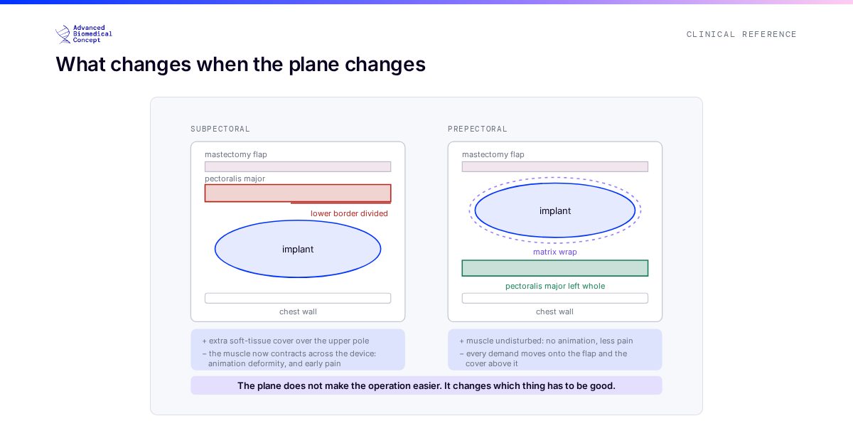
The same implant drawn in two planes. Subpectoral, it sits under the pectoralis major, which has been divided along its lower border: the muscle gives extra soft-tissue cover over the upper pole, but it now contracts across the device, which is the mechanism of animation deformity and of post-operative pain. Prepectoral, the implant sits on the muscle, wrapped in a matrix and covered by the mastectomy flap alone: the muscle is left whole and undisturbed, so there is no animation and less early pain, and in exchange every demand shifts onto the quality of the flap and the soft-tissue cover above it.
Clinical Insights — 10 September 2026

Prepectoral or Subpectoral: What Changes When the Plane Changes

Prepectoral placement moves the burden from the muscle to the mastectomy flap. An improvement where the flap can carry it, a mistake…

Read article →
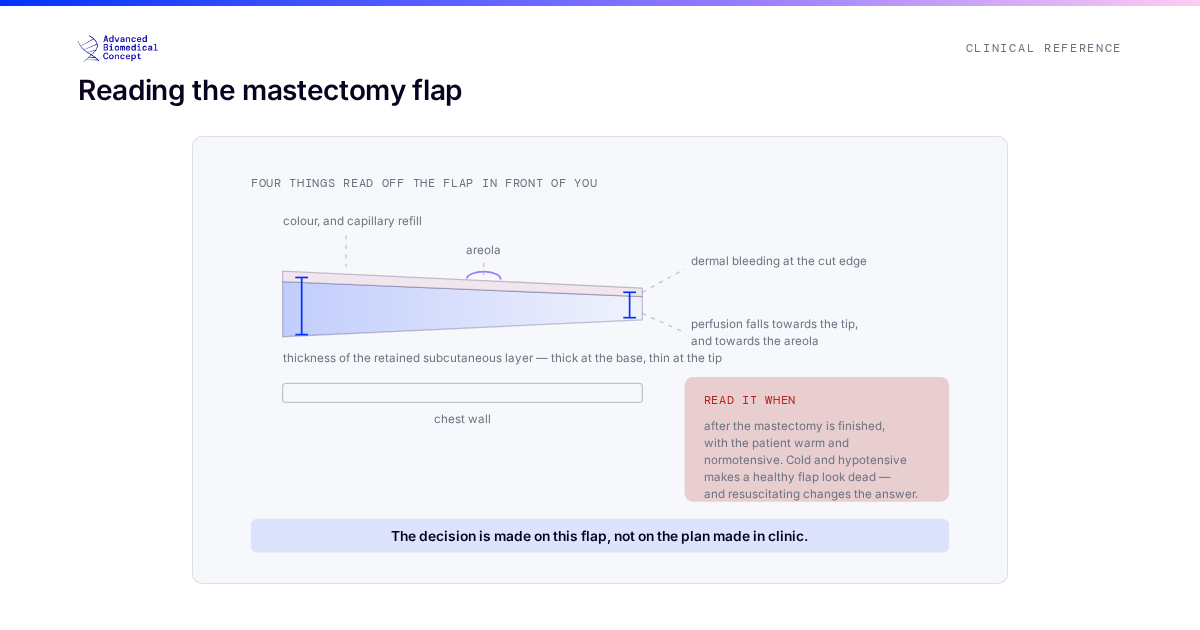
A mastectomy flap in cross-section with the four things assessed before committing to a one-stage reconstruction marked on it: thickness of the retained subcutaneous layer, the colour and capillary refill of the skin, dermal bleeding at the cut edge, and perfusion of the flap as a whole shown as a gradient that falls towards the distal tip and towards the areola. A panel beside it notes that the flap is judged after the mastectomy is finished and under normal blood pressure and temperature, because a cold, hypotensive patient makes a healthy flap look dead and warming and resuscitation change the reading.
Clinical Insights — 07 September 2026

Reading the Mastectomy Flap: The Decision That Is Still Open in Theatre

Direct-to-implant is a single-stage operation resting on a judgement made mid-operation. What is assessed, why it cannot be decided beforehand, and what…

Read article →
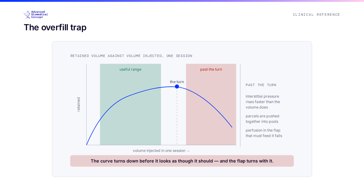
A curve of retained volume plotted against volume injected in a single session. It rises steeply, flattens, and then turns downward. Under the flat part the tissue is accepting fat at low interstitial pressure. Past the turn, pressure rises faster than volume, the parcels are pushed together into pools, perfusion in the bed falls, and both the graft and the flap that carries it are compromised — so injecting more returns less than injecting less. A marked band shows where the useful range sits, well before the peak.
Clinical Insights — 04 September 2026

Planning Fat Grafting Across Sessions: Volume, Staging and the Overfill Trap

Past a certain volume, added fat does not add retained volume — it subtracts it. Why staging is the plan rather than…

Read article →
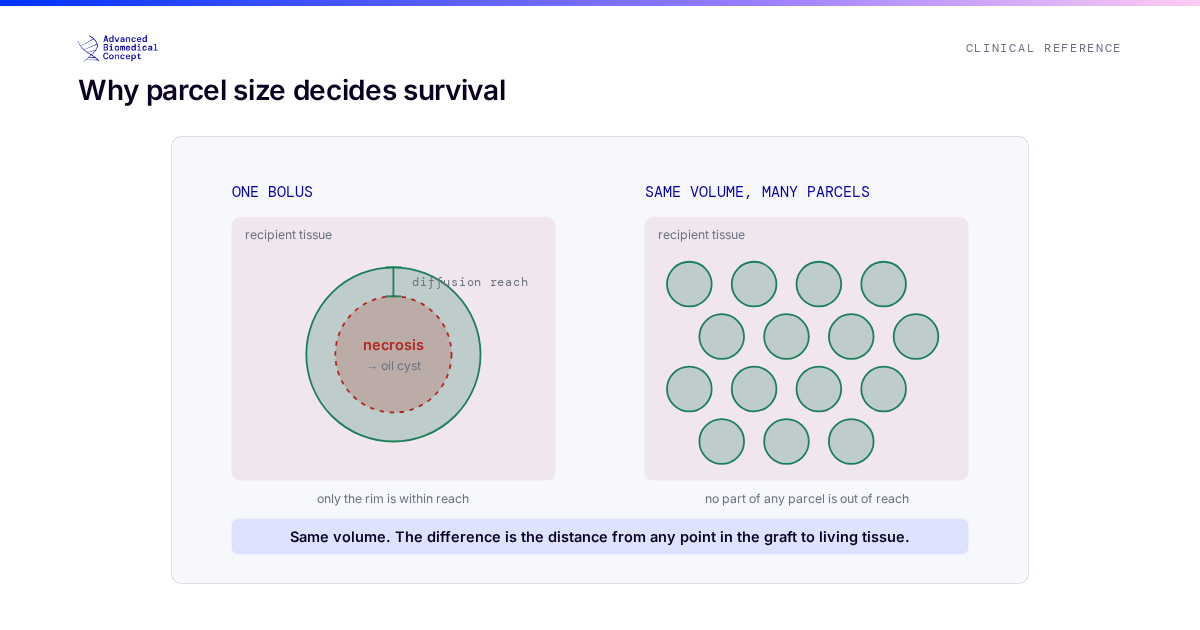
Two grafts drawn in cross-section. The first is a single large bolus: only a thin outer rim lies within reach of the surrounding tissue and survives, while the centre, further away than oxygen can diffuse, becomes necrosis and later an oil cyst. The second is the same total volume placed as many small parcels, each one thin enough that no part of it sits beyond diffusion range, so nearly all of it stays within the surviving zone until new blood vessels arrive.
Clinical Insights — 01 September 2026

What Actually Determines Fat Graft Retention

Retention is not a property of the fat but of the space it was placed into. A surgeon-facing walk through the chain:…

Read article →
questions-to-ask-before-breast-reconstruction-01
Clinical Insights — 21 August 2026

Questions to Ask Your Surgeon Before Breast Reconstruction

Consultations are short and full of unfamiliar vocabulary. The questions worth asking, why each matters, and what a good answer sounds like.

Read article →
fat-grafting-01
Clinical Insights — 21 August 2026

Is Fat Grafting Safe After Breast Cancer? The Oncological Evidence

The laboratory worry, the clinical evidence and the honest caveats: what studies of thousands of patients show about fat grafting after breast…

Read article →
recovery-timeline-01
Clinical Insights — 18 August 2026

Breast Reconstruction Recovery: A Realistic Timeline, Week by Week

Several timelines run at once after reconstruction and they finish at different times. What typically happens, and when, in plain language.

Read article →
hybrid-breast-reconstruction-implant-plus-fat-grafting-01
Clinical Insights — 18 August 2026

Hybrid Breast Reconstruction: Combining an Implant, Matrix Support and Fat Grafting

Implant for structure, fat for the living envelope, matrix for support: the rationale, technique and published outcomes of hybrid reconstruction.

Read article →

Posts navigation

← Newer 1 2 3 … 7 Older →
Advanced Biomedical Concept

Innovators in breast reconstruction surgery. CE-marked biological membranes and surgical support devices. Founded 2020, Rome, Italy.

info@advancedbioconcept.it

Company

  • Our Story
  • Meet the Team
  • Testimonials & Quotes
  • Events
  • Certifications
  • Products

  • Exashape Line
  • Exafat Kit
  • Clinical Guides

    Biological Matrix for Breast Reconstruction Prepectoral Breast Reconstruction Rigottomia (Rigottomy): The Technique Explained Biological Matrix, ADM & Synthetic Mesh: A Clinical Comparison View all clinical guides →

    For Professionals

    Clinical StudiesE-LearningContact

    Tools

  • Decision Simulator
  • Media Kit
  • Exashape Evidence Library
  • Cost-Per-Case Calculator
  • Breast Reconstruction Options ExplorerDRAFT Libra

    © 2026 Advanced Biomedical Concept Srl. All rights reserved. — P.IVA: 15657681001

    Privacy PolicyCookie settings For Healthcare Professionals Only. CE marked per EU MDR 2017/745.
    Advanced Biomedical Concept
    Discover what we make

    From the ExaShape® biological matrix for breast reconstruction to the EXAFAT® fat-grafting system — explore the full range of ABC devices.

    Explore our products →

    We value your privacy We use essential cookies to make our site work. With your consent we may also use cookies to analyse traffic and improve your experience. See our Privacy Policy and Cookie Policy.

    How can we help?

    Send us a message and our team will get back to you within 1–2 business days.
    We typically reply within 1–2 business days.