ExaShape · Structural Explainer
Not all matrices share a biography
Two biological matrices can look identical on the back table and behave differently, because their source tissue shapes their architecture. Toggle between source tissues, then trace the ExaShape bovine pericardium mode of action across twelve months.
For Healthcare Professionals Only
CE · EU MDR 2017/745
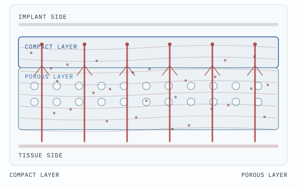
Acellular Bovine Pericardium
Compact layerPorous layer
Bovine pericardium
ExaShape · bilayer by nature
Mode of action, across 12 months
Day 0 · Implantation
Day 0
Inflammation
Neoangiogenesis
Collagen
Remodelling
The timeline above traces the documented ExaShape bovine pericardium mode of action. Characteristics of dermal matrices vary by manufacturer, source, and processing, so no single timeline is shown here. Switch back to bovine pericardium to trace the ExaShape sequence.
This explainer describes structural and biological characteristics of each source tissue for educational purposes. It is not a comparative performance claim between commercial devices. ExaShape data is drawn from the cited real-world clinical studies.
- De Vita R, et al. A Pericardium Bovine Matrix Pocket in DTI Prepectoral Breast Reconstruction. Clinical Breast Cancer. 2024.
- Varvaras D, et al. Use of Acellular Pericardial Biological Mesh for Prepectoral and Dual Plane. St. Gallen 2023.
- Bernardini R, et al. J Biomed Mater Res. 2020.
ExaShape is CE marked per EU MDR 2017/745. For Healthcare Professionals Only. Images and illustrations are schematic and for illustrative purposes only.